UNIVERSITÀ DEGLI STUDI DI PADOVA DIPARTIMENTO DI SCIENZE CHIMICHE SCUOLA DI DOTTORATO DI RICERCA IN SCIENZE MOLECOLARI INDIRIZZO: SCIENZE CHIMICHE XXI CICLO Production and characterization of SulP anion transporters Direttore della Scuola: Ch.mo Prof. Maurizio Casarin Supervisore: Ch.mo Prof. Roberto Battistutta Dottoranda: Elisa Pasqualetto 2 febbraio 2009
Transcript
UNIVERSITÀ DEGLI STUDI DI PADOVA
DIPARTIMENTO DI SCIENZE CHIMICHE
SCUOLA DI DOTTORATO DI RICERCA IN SCIENZE MOLECOLARI
INDIRIZZO: SCIENZE CHIMICHE
XXI CICLO
Production and characterization of SulP anion transporters
Direttore della Scuola: Ch.mo Prof. Maurizio Casarin
Supervisore: Ch.mo Prof. Roberto Battistutta
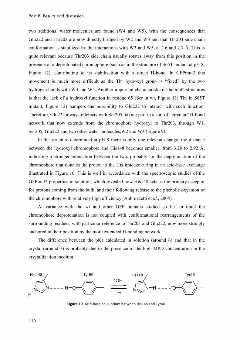
Dottoranda: Elisa Pasqualetto
2 febbraio 2009
Ai miei genitori
I
CCoonntteennttss
Summary 1
Riassunto 3
Part A: Production and characterization of SulP anion transporters
1 Introduction
1.1 The Sulphate Permease (SulP) family 9
The SLC26 gene family 9
The SLC26 family and genetic diseases 10
Membrane topology of the SulP proteins 11
The transmembrane domain 12
The STAS domain 13
The STAS domain and genetic diseases 13
ASA proteins STAS domain 14
Anion transporters STAS domain 15
SULTR1.2 STAS domain 18
Rv1739c STAS domain 19
1.2 The protein prestin 21
OHC electromotility 21
The discovery of prestin 24
Prestin and deafness 26
Reciprocal electromechanical properties of prestin 26
Prestin topology 27
Mechanism of action 28
Incomplete transporter 29
Anion antiporter 30
II
Prestin STAS domain 31
Oligomerization properties 33
Prestin orthologs 35
2 The project
Aims of this study 39
The strategy 39
Production and characterization of the STAS domain 40
Production of SulP proteins by cell-free expression system 43
3 Results and discussion
3.1 Overview 47
3.2 Expression, purification and characterization of prestin STAS domain 49
Experimental procedures 49
Design of three variants of the C-terminal domain of prestin 49
Cloning of prestin genes into the expression vectors 50
Proteins expression 50
Purification and proteolytic cleavage of fusion proteins 51
Analytical reverse phase chromatography and mass spectrometry 51
Circular dichroism (CD) spectroscopy 51
Fluorescence spectroscopy 51
Analytical gel permeation chromatography 52
Dynamic light scattering (DLS) 52
Crystallization tests 52
Results and discussion 52
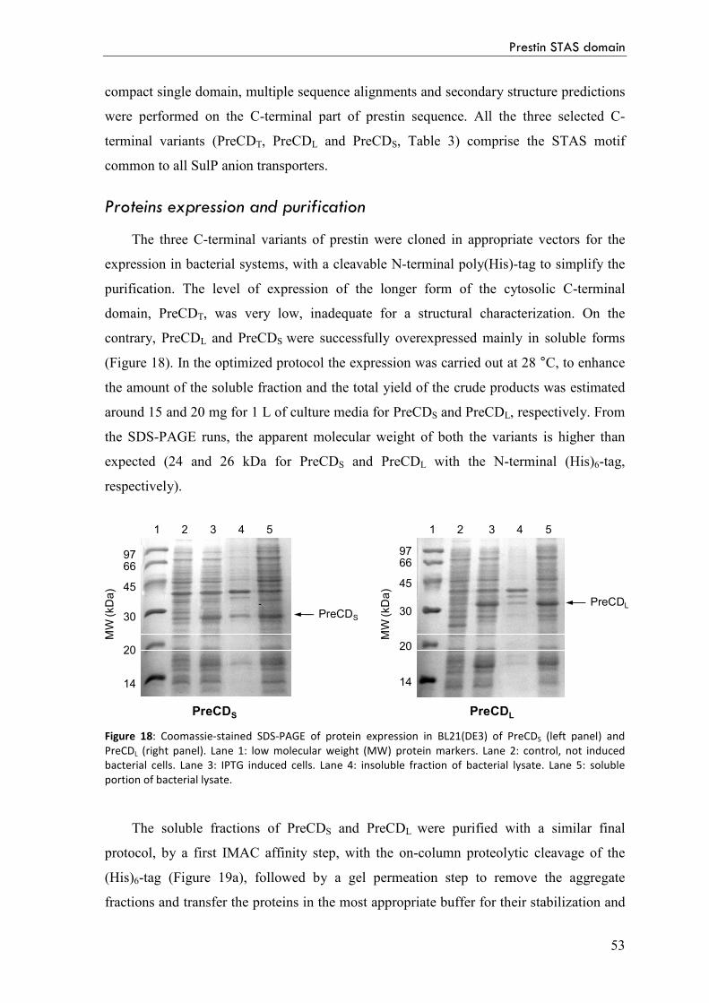
Proteins expression and purification 53
Circular dichroism (CD) and fluorescence spectroscopy 55
Oligomerization properties 56
3.3 Expression, purification and characterization of Rv1739c STAS domain 61
Experimental procedures 61
Design of two variants of the C-terminal domain of Rv1739c 61
Cloning of Rv1739c genes into the pET SUMO expression vector 61
Proteins expression 62
III
Purification and proteolytic cleavage of fusion proteins 62
Analytical reverse phase chromatography and mass spectrometry 63
Analytical gel permeation chromatography 63
Circular dichroism (CD) spectroscopy 63
Crystallization tests 63
Results and discussion 64
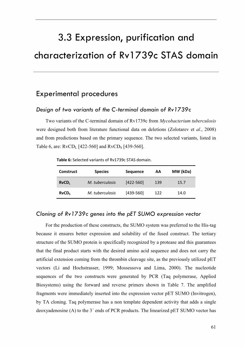
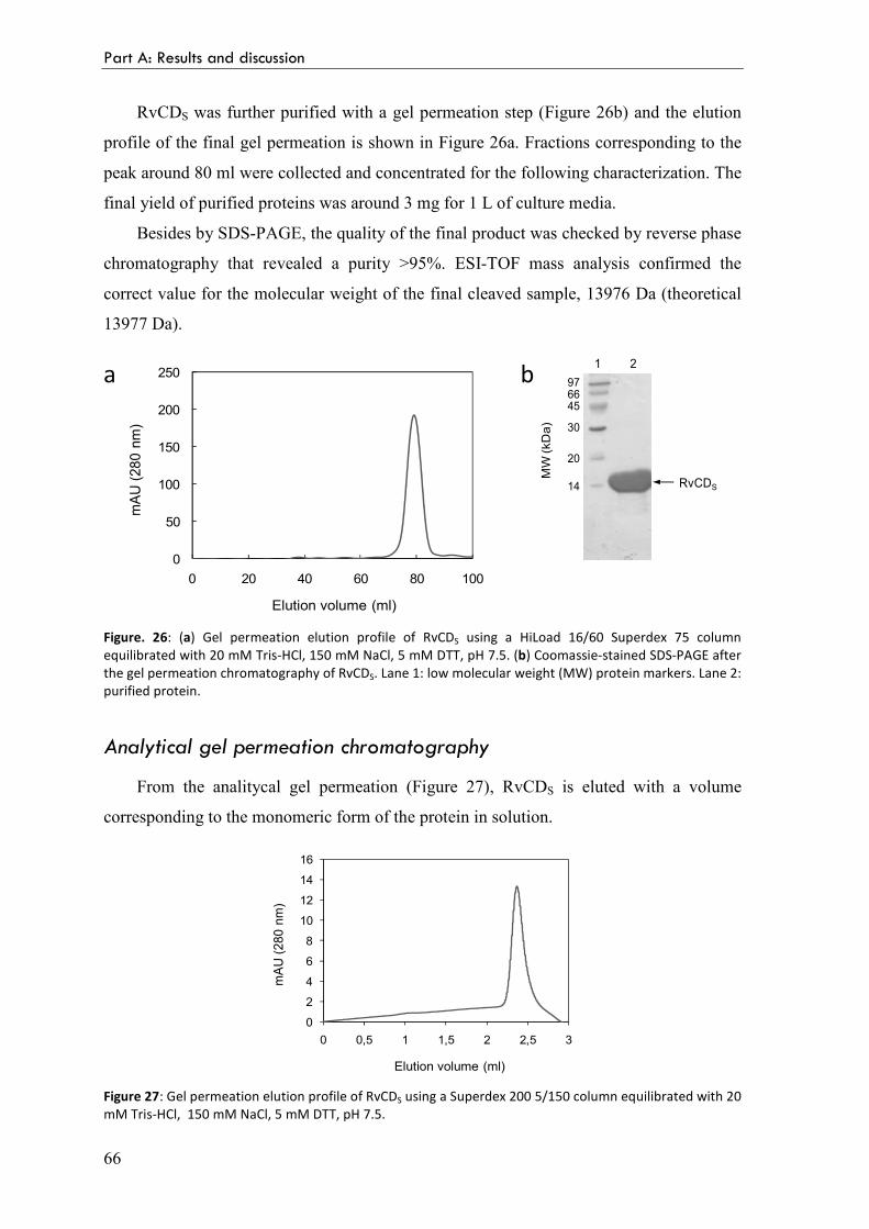
Proteins expression and purification 64
Analytical gel permeation chromatography 66
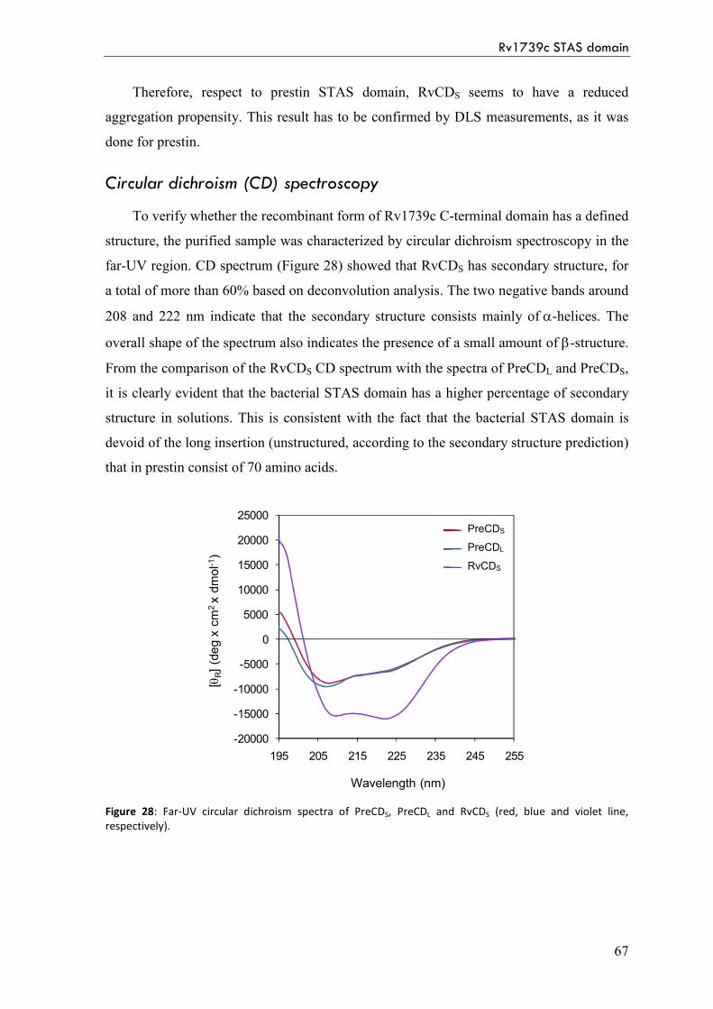
Circular dichroism (CD) spectroscopy 67
3.4 Cell-free expression of full-length SulP proteins 69
Introduction: Cell-free expression of membrane proteins 69
Experimental procedures 71
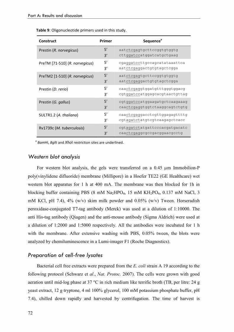
Selection of the SulP proteins for CF expression 71
Cloning of the genes into the pET-21cHX expression vector 71
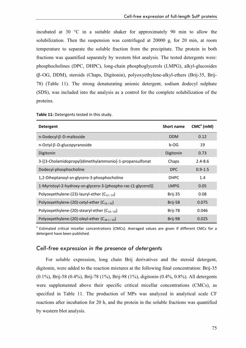
Detergent solubilization of precipitate proteins 74
Cell-free expression in the presence of detergents 75
Results and discussion 76
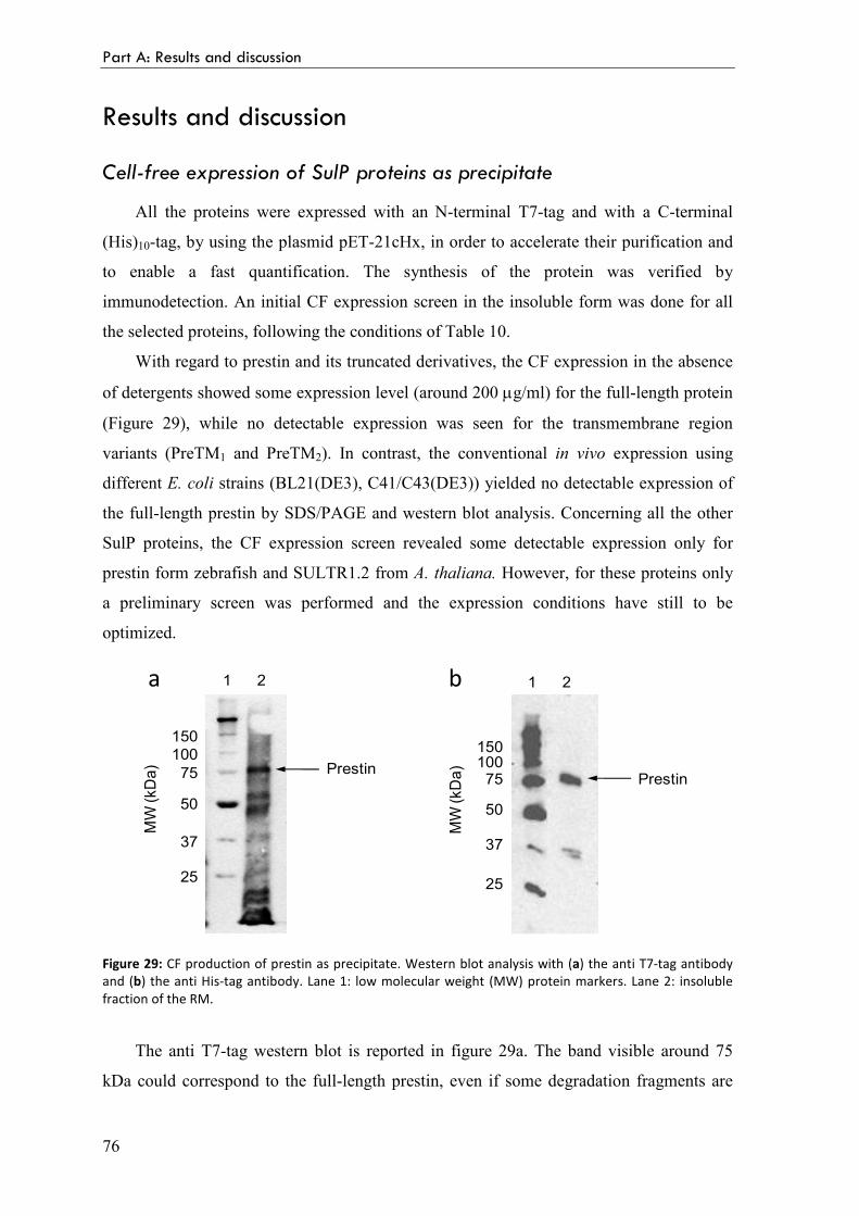
Cell-free expression of SulP proteins as precipitate 76
Optimization of prestin cell-free expression conditions 77
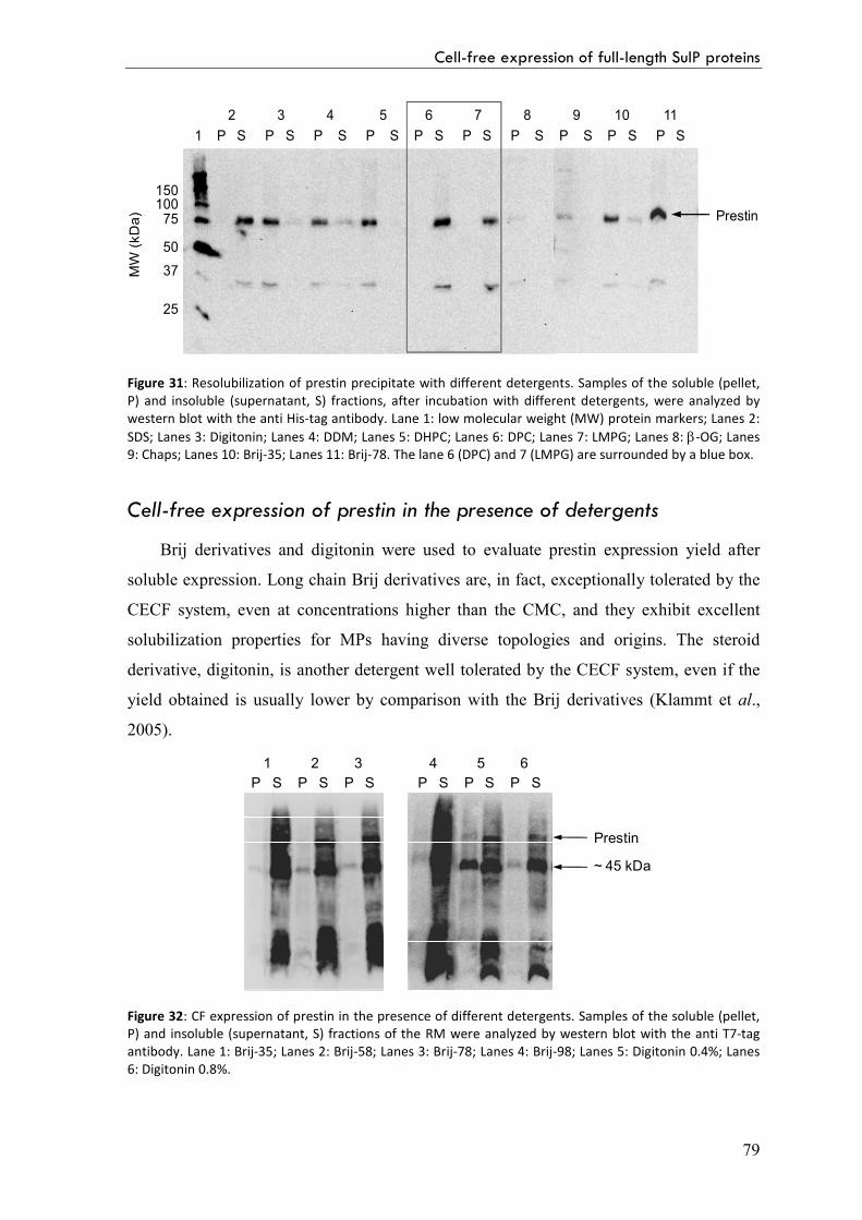
Detergent solubilization of precipitate prestin 78
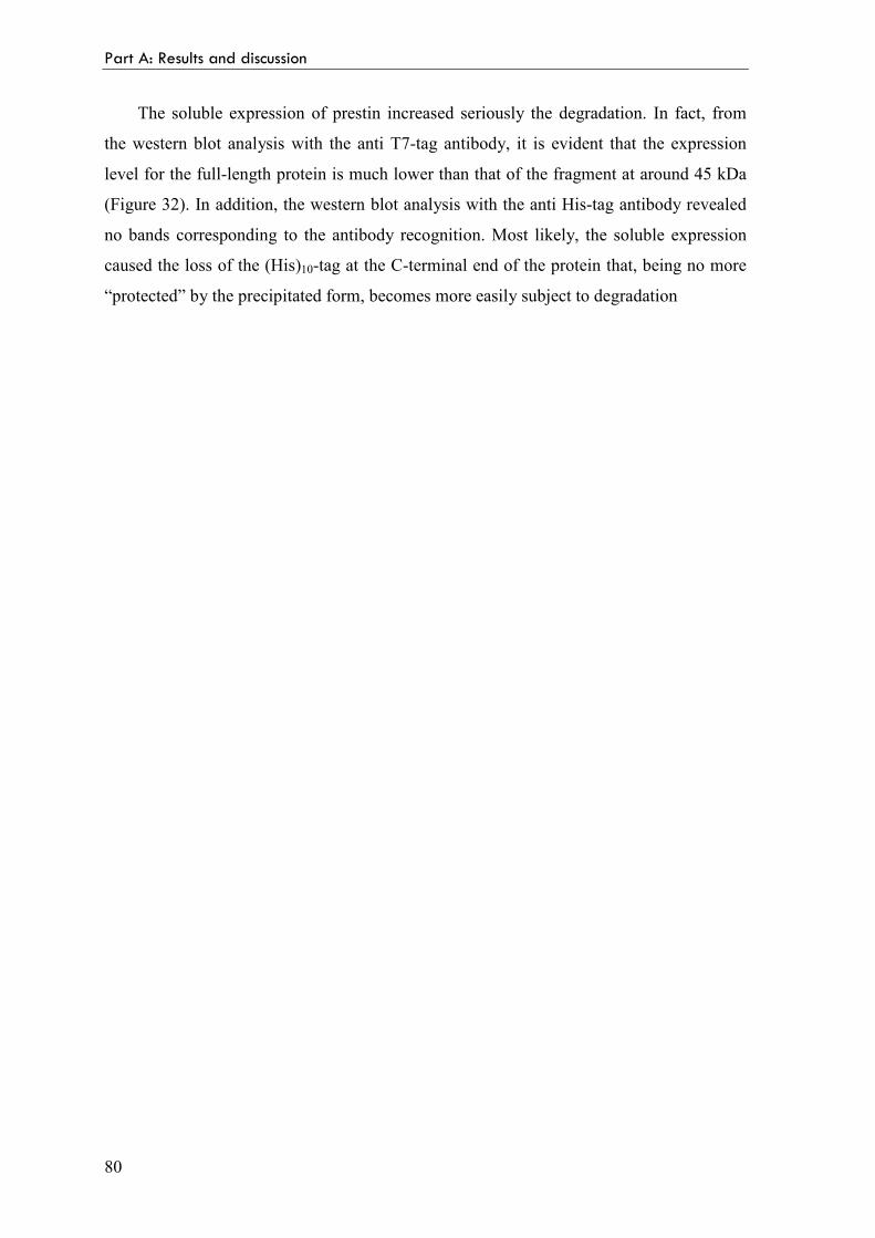
Cell-free expression of prestin in the presence of detergents 79
4 Conclusions 83
References 85
Part B: Structural studies on the Green Fluorescent Protein mutant,
GFPmut2, at different pH
1 Introduction
1.1 The Green Fluorescent Protein (GFP) 99
Crystal structure and chromofore formation 99
IV
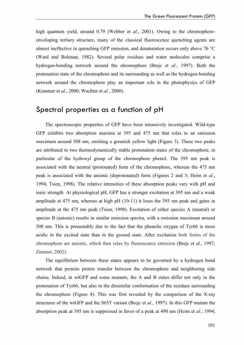
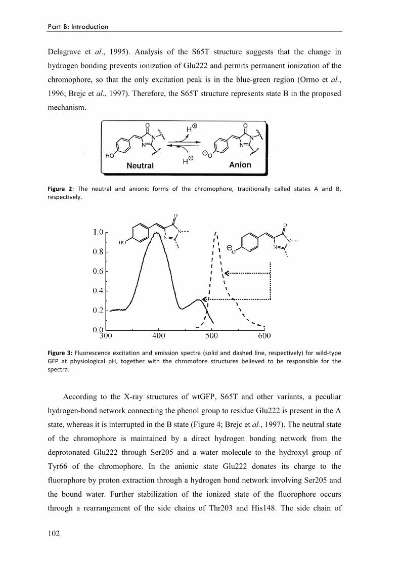
Spectral properties as a function of pH 101
GFP mutants 103
GFPmut2 104
Aim of this study 106
2 Results and discussion
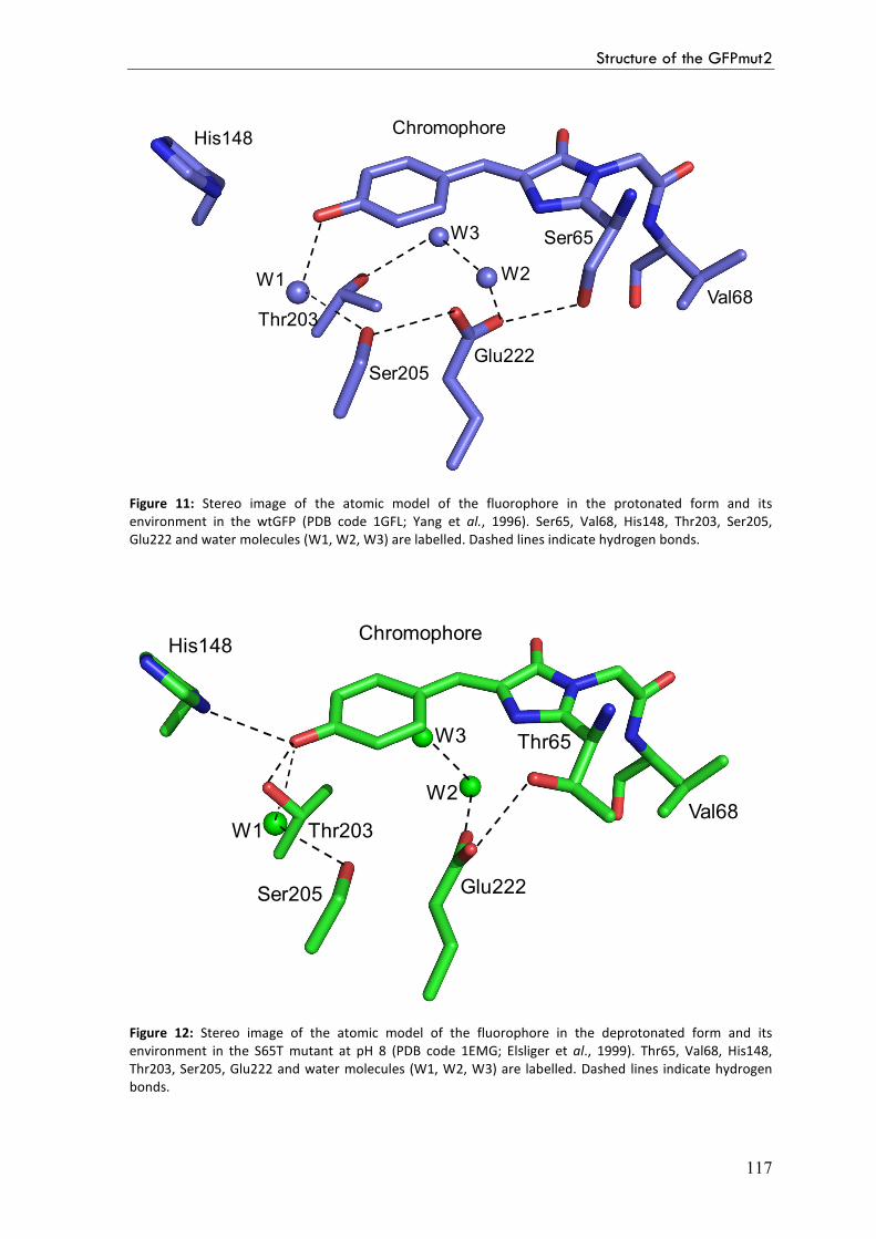
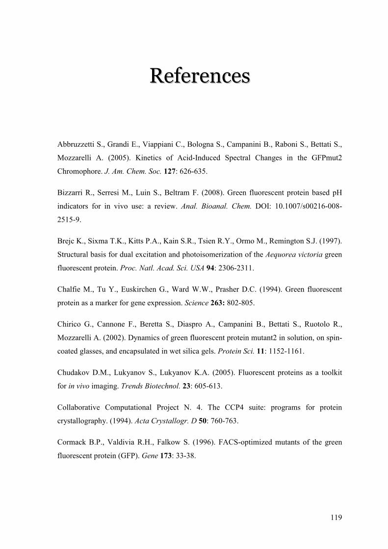
2.1 Structure of the GFPmut2 at both acidic and basic pH 111
Experimental procedures 111
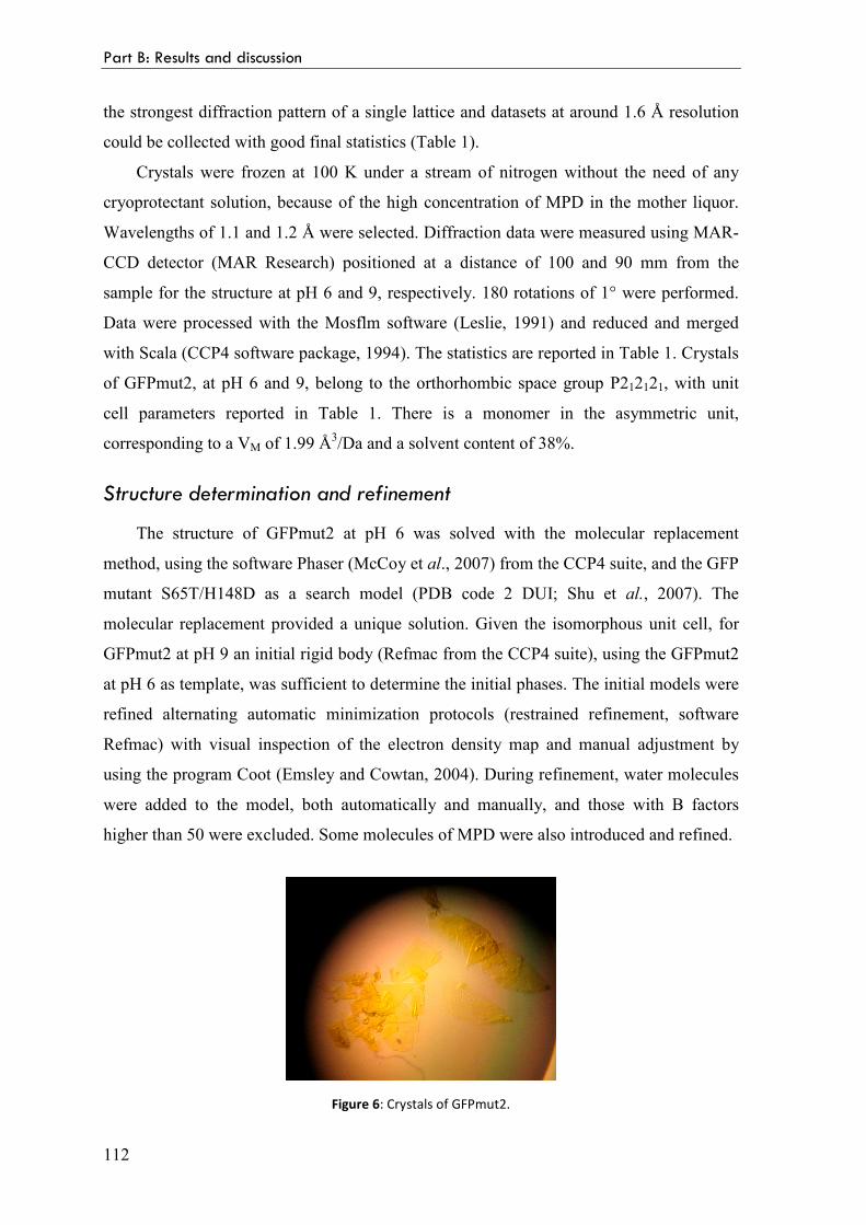
Protein purification and crystallization 111
Spectroscopic analysis 111
Data collection and processing 111
Structure determination and refinement 112
Result and discussion 113
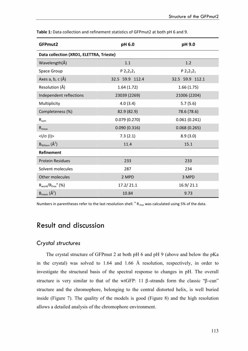
Crystal structures 113
References 119
Abbreviations 123
1
SSuummmmaarryy
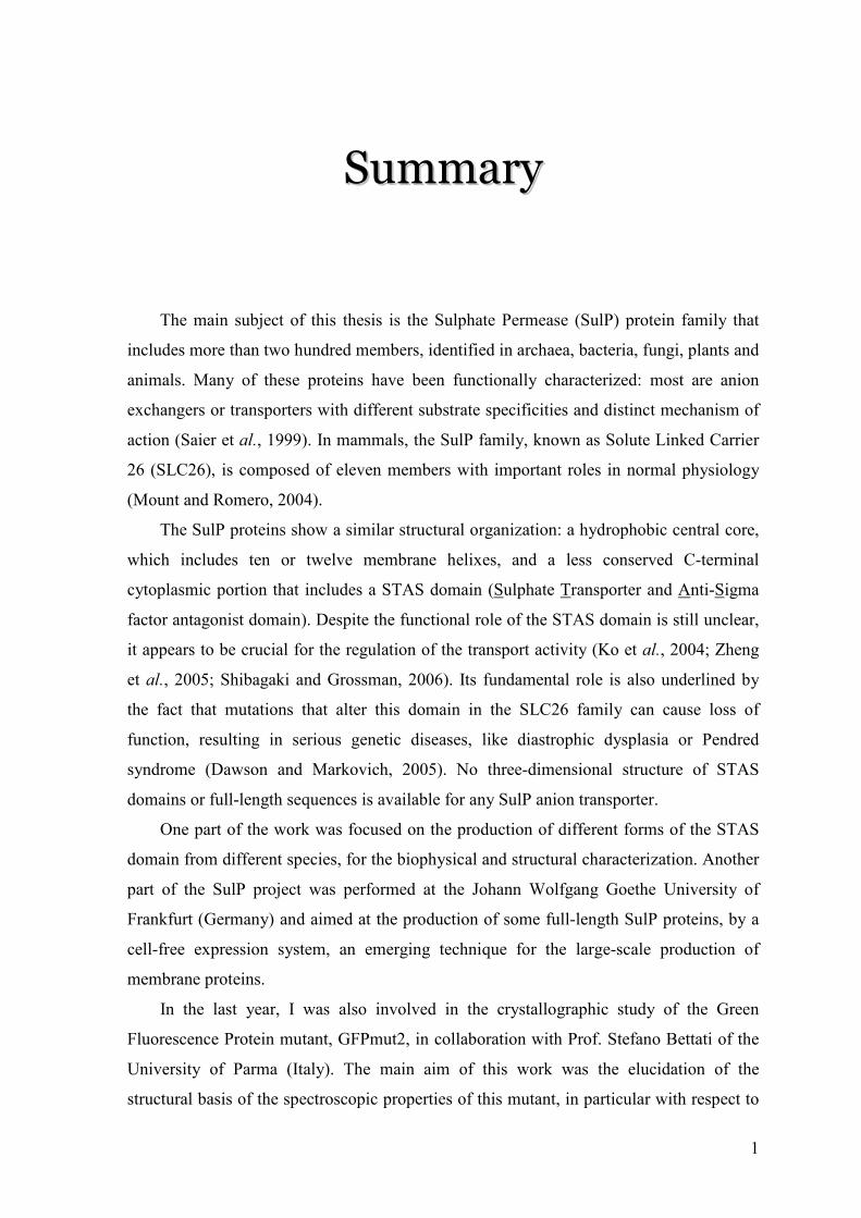
The main subject of this thesis is the Sulphate Permease (SulP) protein family that
includes more than two hundred members, identified in archaea, bacteria, fungi, plants and
animals. Many of these proteins have been functionally characterized: most are anion
exchangers or transporters with different substrate specificities and distinct mechanism of
action (Saier et al., 1999). In mammals, the SulP family, known as Solute Linked Carrier
26 (SLC26), is composed of eleven members with important roles in normal physiology
(Mount and Romero, 2004).
The SulP proteins show a similar structural organization: a hydrophobic central core,
which includes ten or twelve membrane helixes, and a less conserved C-terminal
cytoplasmic portion that includes a STAS domain (Sulphate Transporter and Anti-Sigma
factor antagonist domain). Despite the functional role of the STAS domain is still unclear,
it appears to be crucial for the regulation of the transport activity (Ko et al., 2004; Zheng
et al., 2005; Shibagaki and Grossman, 2006). Its fundamental role is also underlined by
the fact that mutations that alter this domain in the SLC26 family can cause loss of
function, resulting in serious genetic diseases, like diastrophic dysplasia or Pendred
syndrome (Dawson and Markovich, 2005). No three-dimensional structure of STAS
domains or full-length sequences is available for any SulP anion transporter.
One part of the work was focused on the production of different forms of the STAS
domain from different species, for the biophysical and structural characterization. Another
part of the SulP project was performed at the Johann Wolfgang Goethe University of
Frankfurt (Germany) and aimed at the production of some full-length SulP proteins, by a
cell-free expression system, an emerging technique for the large-scale production of
membrane proteins.
In the last year, I was also involved in the crystallographic study of the Green
Fluorescence Protein mutant, GFPmut2, in collaboration with Prof. Stefano Bettati of the
University of Parma (Italy). The main aim of this work was the elucidation of the
structural basis of the spectroscopic properties of this mutant, in particular with respect to
Summary
2
changes in pH. The GFP chromophore can, in fact, exist either in a protonated or
deprotonated state, with distinct spectral properties (Tsien, 1998). In a previous
spectroscopic characterization, GFPmut2 (Ser65Ala, Val68Leu, Ser72Ala) was found
more sensitive than the wild type GFP to pH changes in the physiological range (Chirico
et al., 2002). The structures of GFPmut2 at pH 6 and pH 9 were determined at around 1.6
Å resolution, allowing the correlation between the spectral and structural properties.
3
RRiiaassssuunnttoo
L’oggetto principale di questo lavoro di tesi è la famiglia dei trasportatori anionici
SulP (Sulphate Permease), che comprende più di duecento membri identificati in archea,
batteri, funghi, piante e animali. Molte proteine di questa famiglia sono state
funzionalmente caratterizzate e agiscono da trasportatori o scambiatori di anioni, e
differiscono per l’affinità verso il substrato e il meccanismo di trasporto (Saier et al.,
1999). Nei mammiferi la famiglia SulP, conosciuta come Solute Linked Carrier 26
(SLC26), è composta di undici membri che svolgono un ruolo fondamentale in molti
processi fisiologici nell’uomo (Mount e Romero, 2004).
Tutte le proteine SulP possiedono un’organizzazione strutturale simile: una parte
centrale idrofobica, che comprende dieci o dodici eliche di membrana e una porzione C-
terminale citoplasmatica meno conservata, che include il dominio STAS (Sulphate
Transporter and Anti-Sigma factor antagonist). Sebbene non sia ancora chiaro il ruolo
funzionale di questo dominio nei trasportatori di anioni, esso sembra essere di cruciale
importanza per la regolazione dell’attività di trasporto (Ko et al., 2004; Zheng et al., 2005;
Shibagaki e Grossman, 2006). Il suo ruolo fondamentale è rilevato anche dal fatto che
mutazioni che alterano questo dominio nei membri della famiglia SLC26 possono
comprometterne seriamente la funzionalità, causando malattie genetiche gravi, come la
displasia diastrofica o la sindrome di Pendred (Dawson and Markovich, 2005). Non sono
ancora note strutture tridimensionali di nessun dominio o intera proteina SulP.
Una parte del lavoro è stata focalizzata sulla produzione di diverse varianti del
dominio STAS da specie diverse, finalizzata alla caratterizzazione biofisica e strutturale.
Una seconda parte del progetto, svolta presso la Johann Wolfgang Goethe University di
Francoforte (Germania), ha riguardato la produzione di intere proteine SulP mediante la
sintesi in vitro, una tecnica molto promettente per la produzione su larga scala di proteine
di membrana.
Durante l’ultimo anno, mi sono anche dedicata allo studio cristallografico di un
mutante della Green Fluorescent Protein, GFPmut2, in collaborazione con il gruppo del
Riassunto
4
Prof. Stefano Bettati dell’Università di Parma. L’obiettivo principale di questo lavoro è
stato definire le basi strutturali delle proprietà spettroscopiche di questo mutante, in
particolare al variare del pH. Il cromoforo della GFP può, infatti, esistere sia in forma
protonata che deprotonata (Tsien, 1998). Le proprietà spettroscopiche della GFPmut2
(Ser65Ala, Val68Leu, Ser72Ala) sono state in precedenza caratterizzate e, rispetto alla
proteina wild type, sembra essere più sensibile alle variazioni di pH nell’intervallo
fisiologico (Chirico et al., 2002). A tal fine, è stata determinata la struttura della GFPmut2,
sia a pH 6 che a pH 9, con una risoluzione di circa 1.6 Å. Il confronto delle due strutture
ha consentito la correlazione delle proprietà strutturali con quelle spettroscopiche.
PPaarrtt AA
PPrroodduuccttiioonn aanndd
cchhaarraacctteerriizzaattiioonn ooff
SSuullPP aanniioonn ttrraannssppoorrtteerrss
11
IInnttrroodduuccttiioonn
9
1.1 The Sulphate Permease (SulP) family
The Sulphate Permease (SulP) family is a large and ubiquitous family of membrane
proteins with over two hundred sequenced members, identified by sequence homology in
archaea, bacteria, fungi, plants and animals. Many of these proteins are functionally
characterized: most are anion exchangers (Na+-independent anion transporters) and
transport a wide range of anions, both organic and inorganic, with individual transporters
showing different specificities. Many function by SO42-/H+ symport, but SO4
2-/HCO3-, or
more generally, anion/anion antiport has been reported for several homologues (Saier et
al., 1999).
In bacteria and plants they are responsible for the uptake of sulphate, a convenient
source of sulphur that is a key element in the bacterial as well as in the eukaryotic
metabolism (Kertesz, 2001). In mammals, the SulP family, also known as the Solute
Linked Carrier 26 (SLC26) family of anion transporters, shows broader anion specificity
and more complex functions (Mount and Romero, 2004).
The SLC26 gene family
The SLC26 family is composed of highly versatile anion transporters, with important
roles in normal physiology and human pathophysiology. A partial list of physiological
processes in which the SLC26 exchangers play critical roles includes outer hair cells
(OHCs) electromotility, skeletal development, synthesis of thyroid hormone,
transepithelial Na+-Cl¯ transport, bicarbonate excretion by the distal nephron, and
bicarbonate secretion by the exocrine pancreas (Mount and Romero, 2004).
SLC26A proteins function as anion exchangers or channels in the luminal or apical
membranes of epithelial tissue and are primarily involved in transport of a wide variety of
monovalent and divalent anions. Each member has different anion specificity and
distinctive tissue distribution; some being expressed in most organs and others with more
restricted tissue expression patterns. To date, eleven human SLC26 genes have been
identified, ten of which were shown to encode proteins that transport one or more
Part A: Introduction
10
substrates, including sulphate, chloride, bicarbonate, iodide, oxalate, formate, hydroxyl,
mannose and fructose (Table 1; Mount and Romero, 2004). SLC26A5 (prestin) was
shown to act as the motor protein of cochlear outer hair cells (Zheng et al., 2000). The
SLC26 family thus exhibits an amazing variety of functions, yet the molecular basis of
this diversity is poorly understood.
Table 1: The SLC26 gene family.
Gene Protein name Reported substrate Tissue distribution Disease association
SLC26A1 Sat-1 SO42-
, oxalate Liver, kidney
SLC26A2 DTDST SO42-
, Cl- Widespread Chondrodysplasias
SLC26A3 DRA, CLD SO4
2-, Cl-, HCO3
-, OH-,
oxalate, formate
Intestine, sweat
gland, pancreas,
prostate
Congenital chloride
diarrhea
SLC26A4 Pendrin Cl-, HCO3-, I-, formate
Inner ear, kidney,
thyroid
Pendred syndrome,
deafness (DFNB4)
SLC26A5 Prestin Cl-? Inner ear Deafness?
SLC26A6 CFEX, PAT-1 SO4
2-, Cl-, HCO3
-, OH-,
oxalate, formate Widespread
SLC26A7 None SO42-
, Cl-, oxalate Kidney
SLC26A8 Tat1 SO42-
, Cl-, oxalate Sperm, brain
SLC26A9 None SO42-
, Cl-, oxalate Lung
SLC26A10 None ? Brain
SLC26A11 None SO42-
Widespread
The SLC26 family and genetic diseases
The clinical relevance of the SLC26 gene family was highlighted with the
identification of pathogenetic mutations in four of its genes, namely SLC26A2, A3, A4
and A5 (Table 1). Although these four genes share significant sequence homology and
encode structurally related proteins, they give rise to distinct clinical phenotypes (Dawson
and Markovich, 2005). SLC26A2 is involved in chondrodysplasias that cause skeletal
defects, including clubbed feet, cleft palate, and short limbed dwarfism. Mutations in the
SLC26A3 gene are linked to congenital chloride-losing diarrhea, a disease in which
patients suffer from watery diarrhea containing elevated Cl- concentrations that can prove
The sulphate permease (SulP) family
11
fatal, if left untreated. SLC26A4 is involved in Pendred syndrome, which is the most
common form of syndromic deafness, characterized by congenital sensorineural hearing
loss and thyroid goiter. SLC26A5 encodes a protein, called prestin that is highly and
almost exclusively expressed in the OHCs of the cochlea. The specific expression pattern
of prestin in the OHCs suggests that it is a candidate gene for human deafness. Indeed
SLC26A5 mutations were identified in individuals with non-syndromic deafness,
confirming the physiological role of prestin in human auditory processing (Liu et al.,
2003; Toth et al., 2007).
Given the different anion specificity and the distinct tissue distribution of expression
for each of these genes, it is not surprising that they are associated with markedly different
clinical phenotypes.
Membrane topology of the SulP proteins
With only a few exceptions, the bacterial SulP transporters vary in size from 434 to
573 residues, while the eukaryotic proteins are usually larger, varying from 611 to 893
residues (Saier et al., 1999).
Although the level of amino acid identity between all members of the SulP family is
low, around 25%, hydropathy plots of different members of the family from bacteria to
humans are clearly similar, suggesting structural and functional similarities. Moreover,
blocks of more highly conserved amino acids are present in some transmembrane helices
and some of these are functionally important. This implies that there will be common
features in the transport mechanisms throughout the family (Loughlin et al., 2002).
Prosite motif
Saier motif
STAS domain
outside
inside
91 2 3 4 5 6 7 8 10 11 12
Figure 1: One predicted topology model of the SulP proteins. The position of various conserved motifs and
domains is depicted. The number of transmembrane helices can vary from 10 to 14.
Part A: Introduction
12
The detailed membrane topology of the SulP exchangers has not been determined
experimentally, and prediction programs yield highly divergent models (Figure 1). The
SulP family proteins are predicted to have 10 to 14 transmembrane spanning α-helices,
with intracellular N- and C-termini (Saier et al., 1999; Mount and Romero, 2004).
The transmembrane domain
Much of the homology between SulP exchangers is found within the hydrophobic
core of transmembrane domain. The first two putative transmembrane α-helices show a
significantly higher level of conservation than that observed for the entire protein. This
region includes one of the two “sulphate transporter motifs” that have been used to define
the SulP family (Saier et al., 1999). The first consensus signature extends across putative
helix 2 and comprises 22 amino acids (Prosite, PS01130; Figure 1). Although not all
members of the family conform to the exact consensus sequence, this region contains
several invariant residues that are presumably critical for anion transport. Moreover, an
alignment of eukaryotic family members shows that there are also positions in helix 1 with
high levels of conservation. In addition to conservation of the residue at each position, the
spacing between them, including a short loop between the first two helices, is maintained
throughout the eukaryotic members of the family (Leves et al., 2008). Mutagenesis studies
on these residues were performed on a plant sulphate transporter SHST1, from the tropical
legume Stylosanthes hamata (Shelden et al., 2001; Loughlin et al., 2002; Leves et al.,
2008) and prestin (SLC26A5), a distantly related mammalian member of the SulP family
(Rajagopalan et al., 2006). These studies confirm the predicted importance of conserved
residues in helices 1 and 2 and suggest that function of the SulP members is dependent on
a network of polar and aromatic interactions between these two helices.
The second cluster of invariant residues defined by Saier and colleagues extends
across putative helix 9 (Figure 1; Saier et al., 1999). This helix is somewhat atypical in
that it contains a great number of polar residues. Two conserved residues in this region
(Asn395 and Glu387) were shown to have functional significance in SHST1 (Khurana et
al., 2000; Loughlin et al., 2002). These studies suggest that putative helix 9 may be
important for stability and/or trafficking of SHST1 to the plasma membrane. Moreover,
mutations in the correspondent residues in two members of the SLC26 family result in
serious diseases. A severe dysplasia, achondrogenesis type Ib, can be caused by a
mutation that affects Asn425 in SLC26A2, equivalent to Asn395 in SHST1. Pendred
The sulphate permease (SulP) family
13
syndrome may be the result of the mutation of Glu384 in SLC26A4 (Glu387 in SHST1).
These results indicate that conserved residues between distinct members of the family may
share essential roles in structure or function.
The STAS domain
The less conserved C-terminal cytoplasmic portion of all SulP proteins extends into
the cytoplasm of the cell and includes a so-called STAS domain. The STAS domain
(Sulphate Transporter and Anti-Sigma factor antagonist domain) was identified by the
sequence analysis of proteins with completely different functions (Aravind and Koonin,
2000). This analysis revealed an unexpected, statistically significant similarity between
the carboxy-terminal cytoplasmatic part of SulP transporters (that can vary in length from
around 115 to around 250 amino acids) and the bacterial Anti-Sigma factor Antagonists
ASA, typified by Bacillus subtilis SpoIIAA (117 residues long).
The STAS domain and genetic diseases
The C-terminus is the least conserved region of the protein among different SLC26A
family members, therefore it is most likely to be responsible for each protein specific
function. Although the STAS domain appears to be of crucial importance for the
regulation of transport activity, the functional role of this domain with respect to anion
transporters is still poorly understood. Its fundamental role is underlined by the fact that
mutations that alter this domain in the SLC26 family can cause loss of function, resulting
in serious diseases, like diastrophic dysplasia, Pendred syndrome, and congenital chloride
diarrhea (Dawson and Markovich, 2005). The majority of these mutant proteins has
improper plasmamembrane targeting and loss of some or full function.
It was shown that mutations in the STAS domain of SLC26A3, which functions as a
coupled Cl-/HCO3- exchanger, result in congenital chloride diarrhea, by causing a loss in
wild type levels of functional protein at the plasma membrane. This is probably caused by
at least two distinct mechanisms: misfolding that prevents the mutant transporters from
reaching the native state, and the disruption of important intramolecular interactions
critical to form a well folded and functional transporter (Dorwart et al., 2008).
Muallem and colleagues provided clear evidence for a reciprocal regulation between
the CFTR chloride channel, implicated in cystic fibrosis, and two members of the SLC26
Part A: Introduction
14
family (SLC26A3 and SLC26A6). The interaction is mediated by binding of the
regulatory (R) domain of CFTR to the STAS domain of SLC26A proteins. The interaction
is modulated by PDZ binding scaffold proteins that tether the two transporters into a
multimeric complex with other regulatory proteins (Ko et al., 2004). These findings
provide new insights into the mechanism of bicarbonate and fluid secretion from epithelial
tissues and may lead to better treatments for cystic fibrosis and congenital chloride
diarrhea (Gray, 2004).
ASA proteins STAS domain
The bacterial SpoIIAA protein is a key component of the regulation network involved
in the induction of sporulation in response to nutrient deficiency. The transcription factor
SpoIIAA or Anti-Sigma factor Antagonist (ASA) associates with the complex formed by
the sigma factor and the anti-sigma factor SpoIIAB; this association causes the release of
the sigma factor from SpoIIAB, triggering sporulation-specific transcription (Diederich et
al., 1994; Kroos et al., 1999). SpoIIAB is also a kinase that can phosphorylate and
inactivate SpoIIAA (Duncan et al., 1996).
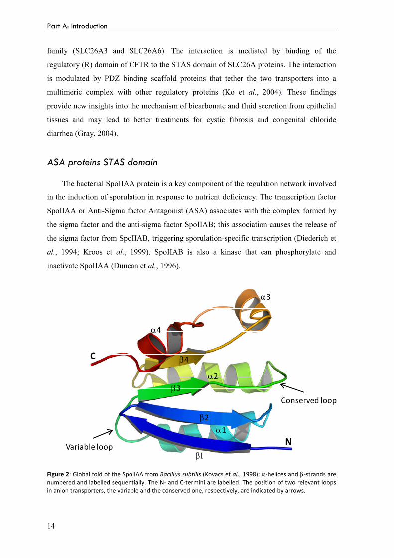
Figure 2: Global fold of the SpoIIAA from Bacillus subtilis (Kovacs et al., 1998); α-helices and β-strands are
numbered and labelled sequentially. The N- and C-termini are labelled. The position of two relevant loops
in anion transporters, the variable and the conserved one, respectively, are indicated by arrows.
α4
α3
α2
α1
β4
β3
β2
β1
C
NVariable loop
Conserved loop
The sulphate permease (SulP) family
15
The bacterial ASA are structurally well characterized in their 3D structure both by
NMR spectroscopy (Kovacs et al., 1998) and X-ray crystallography (Seavers et al., 2001).
The SpoIIAA fold consists of four β-strands, forming a β-sheet, surrounded by four α-
helices (Figure 2). The β-sheet, in association with hydrophobic surfaces of the α-helices,
forms a hydrophobic core that is not readily accessible to the external medium. In contrast,
the peripheral exposed surfaces of α-helices and loops are available for interactions with
molecules in the environment. The carboxy-terminal region forms a characteristic α-
helical handle-like structure.
Anion transporters STAS domain
Unlike the bacterial ASA proteins, the STAS domains present in anion transporters
are poorly characterized in terms of both their function and structure; indeed no 3D
structure of such domains is known yet.
The STAS domain of anion transporters shows low overall sequence identity with
SpoIIAA (about 15-20%). The conservation was traced largely to the four strands that
form the scaffold of the STAS domain. In addition, the turn between the two amino-
terminal strands and the long loop between strand β3 and helix α2 are strongly conserved
in almost all the STAS domains (Figures 2 and 3). This loop and β-pleated sheet were
proposed to play a role in nucleotide binding and hydrolysis, by extension from the known
biochemistry of the anti-sigma factor antagonists (Aravind and Koonin, 2000). It was
shown that SpoIIAA binds GTP and ATP and possesses a weak NTPase activity that is
abolished by phosphorylation or by mutation of the phosphorylable serine in the
conserved loop (Najafi et al., 1996). The strong conservation of this loop in the STAS
domains suggests that it could possess general NTP-binding activity. The conserved loop
is probably involved in phosphate binding and the β-sheet scaffold could accommodate
the rest of the NTP molecule. The presence of a predicted NTP-binding domain in the
cytoplasmic portions of anion transporters indicates that anion transport could be regulated
by intracellular concentrations of GTP and/or ATP.
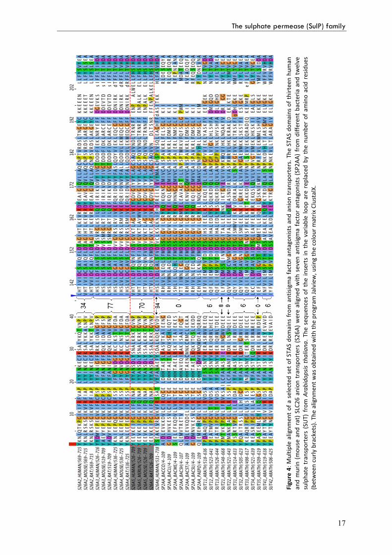
Most of the variability is in the loop between helix α1 and strand β3 (Figures 2 and
3), with inserts of considerable size in some of the anion transporters, of as much as 150
amino acids in the case of SLC26A8 (Aravind and Koonin, 2000). This is immediately
apparent from sequence alignment of all the anion transporters STAS domains and their
Part A: Introduction
16
structural homologs, the SpoIIAA proteins (Figure 4). In the STAS domain of the bacterial
transporters the loop is absent, in the plant sulphate transporter SULTR1.2 it comprises
around 10 residues, while for the mammalian transporters this loop is invariably longer
(for instance around 70 residues for prestin and 150 for SLC26A8). Secondary structure
predictions of this region suggest it is largely unstructured (Dorwart et al., 2008).
Furthermore, in the transporters, a variable extension at the C-terminal end of the domain
is present and the secondary structure predictions of the extreme N- and C-termini do not
correlate with that found in the bacterial ASA.
β-sheet
α-helix
Variable loop
(0-150 amino acids)
Conserved loop
β1 β2 α1 β3 α2 β4 α3 α4
The STAS domain
Figure 3: Representation of the secondary structure elements of the anion transporters STAS domain. A
highly conserved loop is interspersed between strand β3 and helix α2. The STAS domain also contains a
highly variable loop between helix α1 and strand β3. This variable loop is the site of significant insertions in
the SLC26A proteins, of as much as 150 amino acids in the case of SLC26A8.
Taking into account these differences in lengths as well as the low amino acid
conservation observed, most probably the 3D structure of the anion transporters STAS
domains significantly deviates from that of the bacterial ASA, in a way not predictable
solely on the basis of the sequence alignment. Presumably, these differences are
responsible for the distinct properties of this domain when part of the different
transporters.
During the last years, numerous mutagenesis studies were performed on the STAS
domains of different SulP transporters, to elucidate their precise function in the transport
activity. In the following section, two of these studies, concerning a plant and a bacterial
transporter, will be shortly introduced.
The sulphate permease (SulP) family
17
Fig
ure
4:
Mu
ltip
le a
lig
nm
en
t o
f a
se
lect
ed
se
t o
f S
TA
S d
om
ain
s fr
om
an
tisi
gm
a f
act
or
an
tag
on
ists
an
d a
nio
n t
ran
spo
rte
rs.
Th
e S
TA
S d
om
ain
s o
f th
irte
en
hu
ma
n
an
d m
uri
n (
mo
use
an
d r
at)
SLC
26
an
ion
tra
nsp
ort
ers
(S
26
A)
we
re a
lig
ne
d w
ith
se
ve
n a
nti
sig
ma
fa
cto
r a
nta
go
nis
ts (
SP
2A
A)
fro
m d
iffe
ren
t b
act
eri
a a
nd
tw
elv
e
sulp
ha
te t
ran
spo
rte
rs (
SU
T)
fro
m A
rab
ido
psi
s th
ali
an
a.
Th
e s
eq
ue
nce
s o
f th
e i
nse
rts
in t
he
va
ria
ble
lo
op
are
re
pla
ced
by
th
e n
um
be
r o
f a
min
o a
cid
re
sid
ue
s
(be
twe
en
cu
rly
bra
cke
ts).
Th
e a
lig
nm
en
t w
as
ob
tain
ed
wit
h t
he
pro
gra
m J
alv
iew
, u
sin
g t
he
co
lou
r m
atr
ix C
lust
alX
.
Part A: Introduction
18
SULTR1.2 STAS domain
Studies on a sulphate transporter from Arabidopsis thaliana, SULTR1.2, examined
the effect of deleting or modifying the STAS domain. The results suggest that the STAS
domain is essential for facilitating localization of the transporter to the plasma membrane,
but it is also critical for the whole sulphate transport activity (Shibagaki and Grossman,
2004).
The STAS domain of the plant sulphate transporter SULTR1.2 was modeled on the
basis of the available NMR structure of B. subtilis SpoIIAA (1AUZ; Kovacs et al., 1998)
and the crystal structure of B. sphaericus SpoIIAA (1H4Z; Seavers et al., 2001). The
structural analysis and modeling suggest that the SULTR1.2 C-terminal STAS domain
shares the SpoIIAA fold, although it shows low overall sequence identity with SpoIIAA,
around 17% over 130 residues (Figure 5; Rouached et al., 2005). The analysis reveals a
compact hydrophobic core at the interface of the α-helices and the β-sheets. This
hydrophobic core appears very well conserved between the SULTR1.2 STAS domains
and SpoIIAA. The similarity is particularly high in the vicinity of the phosphorylation site,
despite the change from a conserved serine (Ser58) in SpoIIAA to the similar amino acid
threonine (Thr587) in SULTR1.2.
Figure 5: The three-dimensional model of the SULTR1.2 STAS domain. The crystal structure PDB 1H4Z and
the deduced model of the SULTR1.2 STAS domain are shown in red and blue ribbons, respectively. The side
chain of Thr587 (Ser58 in PDB 1H4Z) is represented in green and the two cysteines (Cys645 and Cys646) of
the SULTR1.2 STAS domain are in yellow. α-helices are numbered and labelled sequentially. β-Strands are
not labelled for clarity. The C-terminus of the peptide is labelled, whereas its N-terminus is hidden (behind
helix α2) by the core of the structure (Rouached et al., 2005).
The sulphate permease (SulP) family
19
The major difference between the modeled STAS domain and the SpoIIAA structure
lies at the connection between the SULTR1.2 helix α1 and strand β3 (Figure 4). For the
STAS domain of SULTR1.2, the variable loop comprises around 10 residues. This
variable region lies at the periphery of the domain and is far away from the common
phosphorylation region. The modeled STAS domain also differs from the SpoIIAA
structure at the very C-terminus. This region is highly variable in length and sequence
even between the various sulphate transporters. At the end of the C-terminal helix α4 of
its STAS domain, SULTR1.2 possesses a pair of cysteines (Cys645, Cys646) that are not
strictly conserved in the paralogs and that are not present in SpoIIAA (Figure 5). The two
cysteins seem to play a critical role to maintain the full functionality of SULTR1.2
(Rouached et al., 2005).
An experiment of random mutagenesis in the STAS domain of SULTR1.2 identified
domain lesions that altered the transporter biogenesis and/or function (Shibagaki and
Grossman, 2006). A number of mutations in the β-sheet that forms the core of the STAS
domain prevent plasmamembrane accumulation of SULTR1.2. So the β-sheet seems to
serve as a core structure of the STAS domain and lesions within this structure may disrupt
proper STAS packing, which could destabilize the entire transporter. In contrast, the N-
termini of the first and second α-helices have a number of amino acids critical for the
function of the protein; mutations in these regions still allow protein accumulation in the
plasmamembrane, but the protein is no longer capable of efficiently transporting sulphate
into cells. These results confirm the critical role of the STAS domain for both the activity
and biosynthesis/stability of the transporter, and that defined portions of the STAS domain
correlate with these specific functions.
Rv1739c STAS domain
The SulP family members have been minimally characterized in bacteria. Anyway, it
has been recently shown that induction of Rv1739c expression in E. coli increases
bacterial uptake of sulphate (Zolotarev et al., 2008).
Sulphate uptake was also increased by overexpression of the Rv1739c
transmembrane domain, but not of the cytoplasmic C-terminal STAS domain [437-560].
Expression of the isolated C-terminal cytoplasmic domain did not affect sulphate uptake.
So, unlike the STAS domain requirement for sulphate transport by A. thaliana SULTR1.2
Part A: Introduction
20
(Shibagaki and Grossman, 2004), the STAS domain was dispensable for the sulphate
uptake by Rv1739c.
21
1.2 The protein prestin
Prestin is the fifth member (A5) of the Solute Linked Carrier 26 (SLC26) family of
anion exchangers. It is highly and almost exclusively expressed in the outer hair cells
(OHCs) of the organ of Corti in the inner ear of mammals. Although the basic function of
SLC26A members is to transport anions (Mount and Romero, 2004), this is not prestin
principal role. Unlike the other members of the SLC26 family, mammalian prestin has the
unique property of the voltage-dependent conformational changes and it is considered the
key player in the OHCs somatic electromotility (Zheng et al., 2000). Since its discovery, it
was clear that prestin is fundamentally different from other biological force generators. Its
potential nanotechnology applications make it the most interesting subject among
SLC26A family members, as shown by the increasing number of publications within
recent years. For these reasons, I decided to deal with prestin separately in this section.
OHC electromotility
Cochlear hair cells are non-neuronal epithelial cells that transduce acoustic signals.
They are organized in a tonotopic fashion, with those sensitive to high-pitched sounds at
the basal end and those sensitive to low pitches at the apical end (Géléoc and Holt, 2003).
Perpendicular to this gradient there are four rows of cells: a single row of inner hair cells,
and three rows of outer hair cells (Figure 6). The inner hair cells (IHCs) transduce and
transmit auditory information to the brain. Outer hair cells (OHCs) provide local
mechanical amplification in the form of feedback, thus amplifying the auditory stimuli
sensed by the inner hair cells (Dallos, 1992).
In practice, a pure tone stimulus causes the passive basilar membrane of the organ of
Corti to resonate at a unique location that depends on frequency (Figure 8a). Active
feedback refines or tunes the resonant location and amplifies the membrane motion,
thereby enhancing auditory sensitivity to faint sounds by more than 40 decibel (that is 100
fold) (Dallos, 1992). There is a great deal of evidence indicating that OHCs are the
principal players providing the feedback that drives cochlear amplification.
Part A: Introduction
22
Figure 6: A cross section of the cochlea illustrating the organ of Corti, the sensory epithelium of the inner ear. A single row of inner hair cells and three rows of outer hair cells are located on the basilar membrane. The tectorial membrane overlies the epithelium and normally contacts the stereocilia of the outer hair cells (Dallos and Fakler, 2002).
For the physiological mechanism of amplification two candidate mechanisms have
been proposed. One proposal, for which there is evidence in non-mammalian species, is
that the apical stereocilia of OHCs act both as the sensors of the motion of the basilar
membrane and as a motor source to amplify the motion (Hudspeth et al., 2000). An
alternative theory is based on the motility of the mammalian OHCs. OHCs have a
distinctive hair (stereocilia) bundle (Figure 7), which is the mechanosensory input
organelle of these cells. When mechanically stimulated by incoming sound waves, the
ciliary bundle is deflected, and thereby triggers the opening and closing of
mechanosensitive ion channels in the
stereocilia membrane (Flock et al., 1962;
Hudspeth and Corey, 1977). But, unlike all
other hair cells, OHCs then translate the
resulting changes in membrane potential into
macroscopic changes (up to 5%) in the length
of their cylindrical cell bodies (Evans and
Dallos, 1993). Depolarization triggers cell
contraction, whereas hyperpolarization results
in cell elongation (Brownell et al., 1985;
Kachar et al., 1986). This “electromotility”
Figure 7: Electron microscopy image of the bundle of the stereocilia on the apical surface of outer hair cell in adult mouse (www.neuroscience.cam.ac.uk).
The protein prestin
23
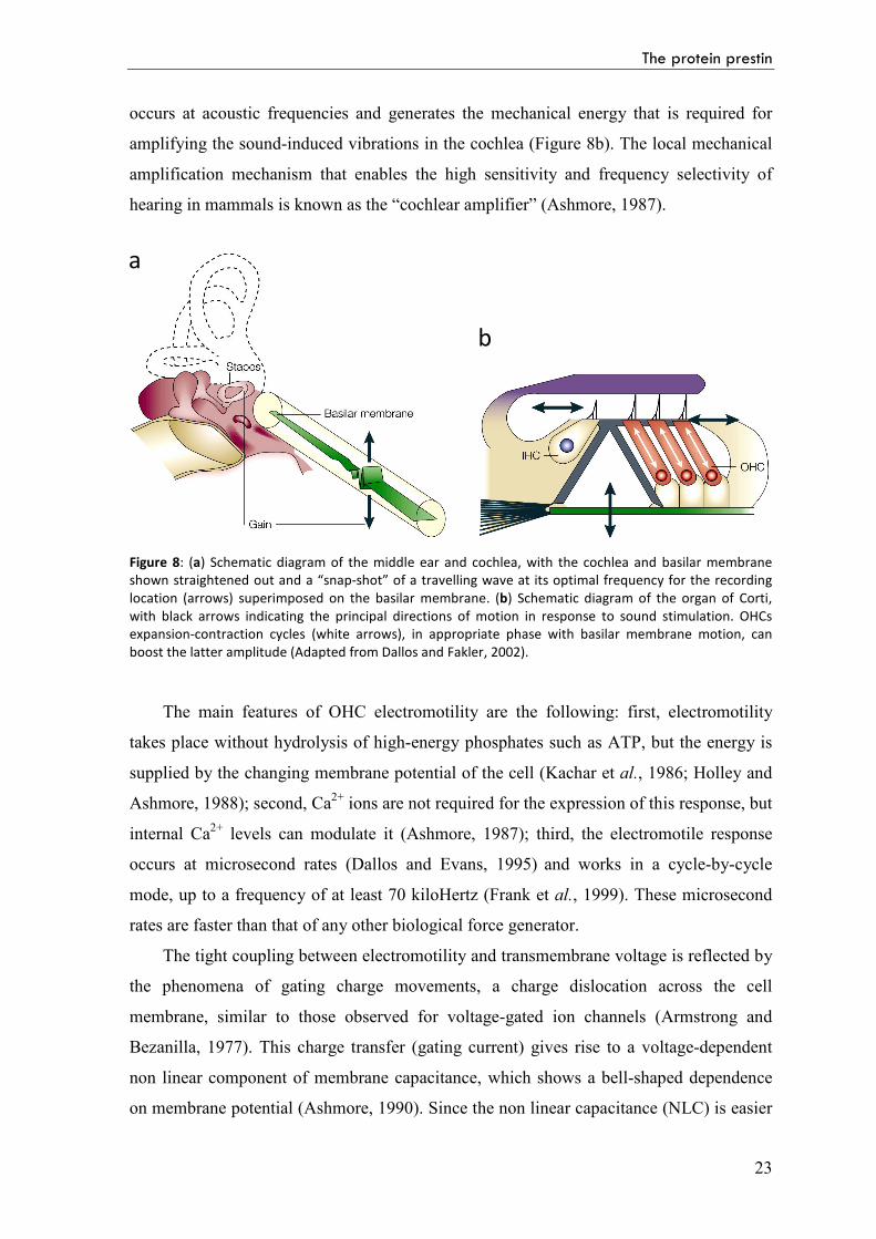
occurs at acoustic frequencies and generates the mechanical energy that is required for
amplifying the sound-induced vibrations in the cochlea (Figure 8b). The local mechanical
amplification mechanism that enables the high sensitivity and frequency selectivity of
hearing in mammals is known as the “cochlear amplifier” (Ashmore, 1987).
Figure 8: (a) Schematic diagram of the middle ear and cochlea, with the cochlea and basilar membrane shown straightened out and a “snap-shot” of a travelling wave at its optimal frequency for the recording location (arrows) superimposed on the basilar membrane. (b) Schematic diagram of the organ of Corti, with black arrows indicating the principal directions of motion in response to sound stimulation. OHCs expansion-contraction cycles (white arrows), in appropriate phase with basilar membrane motion, can boost the latter amplitude (Adapted from Dallos and Fakler, 2002).
The main features of OHC electromotility are the following: first, electromotility
takes place without hydrolysis of high-energy phosphates such as ATP, but the energy is
supplied by the changing membrane potential of the cell (Kachar et al., 1986; Holley and
Ashmore, 1988); second, Ca2+ ions are not required for the expression of this response, but
internal Ca2+ levels can modulate it (Ashmore, 1987); third, the electromotile response
occurs at microsecond rates (Dallos and Evans, 1995) and works in a cycle-by-cycle
mode, up to a frequency of at least 70 kiloHertz (Frank et al., 1999). These microsecond
rates are faster than that of any other biological force generator.
The tight coupling between electromotility and transmembrane voltage is reflected by
the phenomena of gating charge movements, a charge dislocation across the cell
membrane, similar to those observed for voltage-gated ion channels (Armstrong and
Bezanilla, 1977). This charge transfer (gating current) gives rise to a voltage-dependent
non linear component of membrane capacitance, which shows a bell-shaped dependence
on membrane potential (Ashmore, 1990). Since the non linear capacitance (NLC) is easier
a
b
Part A: Introduction
24
to measure than motility, it is widely used as a signature of the electromotile process
(Santos-Sacchi, 1991).
As a consequence of all these observations, it is reasonable to assume that the fast
mechanical changes in OHCs are powered by a molecular motor that is fundamentally
different from other biological force generators, such as the myosin, kinesin or dynein
families. The OHC molecular motor performs direct, rapid, reversible electro-mechanical
conversion (Zheng et al., 2000).
The discovery of prestin
All these findings led to the hypothesis of an integral membrane protein, termed the
motor protein, as the molecular element underlying fast OHC motility (Dallos et al. 1991;
Kalinec et al., 1992). In response to changes in the transmembrane voltage, the motor
protein is thought to undergo a structural rearrangement that changes its area in the plasma
membrane (Dallos et al., 1993; Iwasa, 1994). As a result of the concerted action of a large
number of motor molecules supposed to be densely packed in the OHCs basolateral
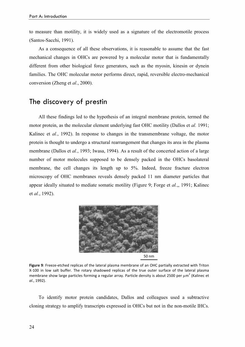
membrane, the cell changes its length up to 5%. Indeed, freeze fracture electron
microscopy of OHC membranes reveals densely packed 11 nm diameter particles that
appear ideally situated to mediate somatic motility (Figure 9; Forge et al.,, 1991; Kalinec
et al., 1992).
50 nm
Figure 9: Freeze-etched replicas of the lateral plasma membrane of an OHC partially extracted with Triton X-100 in low salt buffer. The rotary shadowed replicas of the true outer surface of the lateral plasma membrane show large particles forming a regular array. Particle density is about 2500 per µm2 (Kalinec et al., 1992).
To identify motor protein candidates, Dallos and colleagues used a subtractive
cloning strategy to amplify transcripts expressed in OHCs but not in the non-motile IHCs.
The protein prestin
25
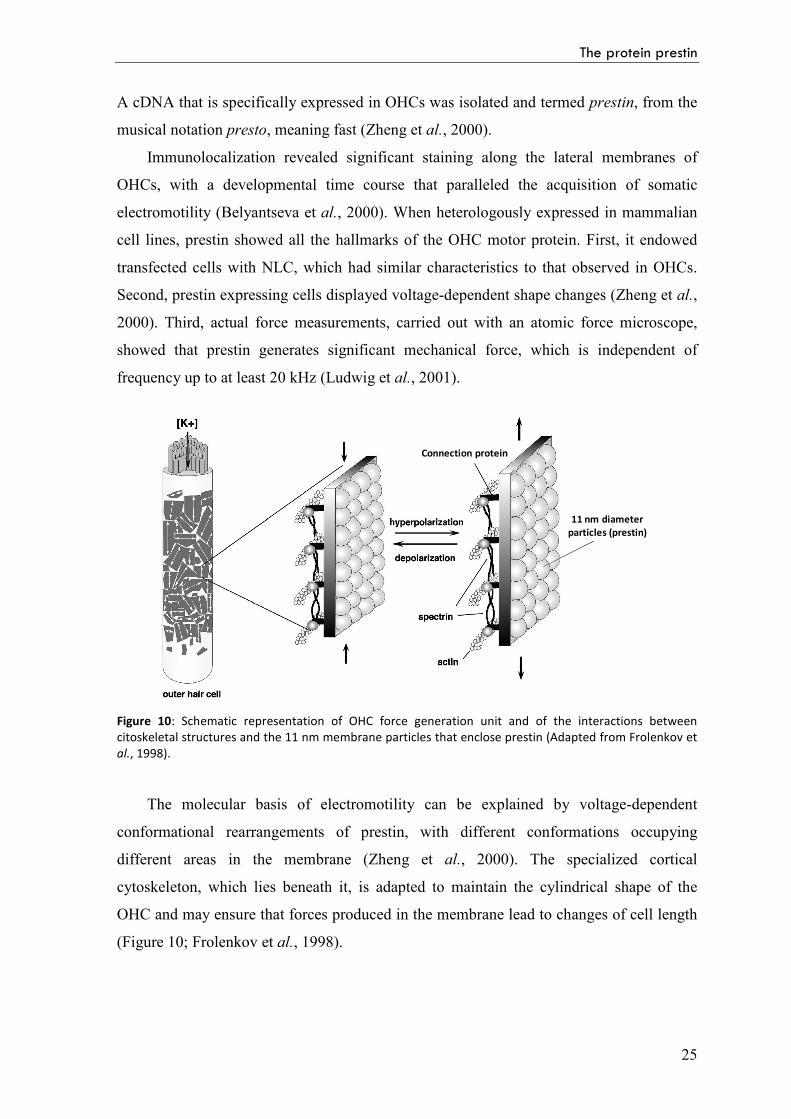
A cDNA that is specifically expressed in OHCs was isolated and termed prestin, from the
musical notation presto, meaning fast (Zheng et al., 2000).
Immunolocalization revealed significant staining along the lateral membranes of
OHCs, with a developmental time course that paralleled the acquisition of somatic
electromotility (Belyantseva et al., 2000). When heterologously expressed in mammalian
cell lines, prestin showed all the hallmarks of the OHC motor protein. First, it endowed
transfected cells with NLC, which had similar characteristics to that observed in OHCs.
2000). Third, actual force measurements, carried out with an atomic force microscope,
showed that prestin generates significant mechanical force, which is independent of
frequency up to at least 20 kHz (Ludwig et al., 2001).
Connection protein
11 nm diameter
particles (prestin)
Figure 10: Schematic representation of OHC force generation unit and of the interactions between citoskeletal structures and the 11 nm membrane particles that enclose prestin (Adapted from Frolenkov et al., 1998).
The molecular basis of electromotility can be explained by voltage-dependent
conformational rearrangements of prestin, with different conformations occupying
different areas in the membrane (Zheng et al., 2000). The specialized cortical
cytoskeleton, which lies beneath it, is adapted to maintain the cylindrical shape of the
OHC and may ensure that forces produced in the membrane lead to changes of cell length
(Figure 10; Frolenkov et al., 1998).
Part A: Introduction
26
Prestin and deafness
The restricted expression of prestin in OHCs and its proposed function as a
mechanical amplifier make it a strong candidate for an association with human deafness.
However, the role and the extent of the prestin gene defects in human non-syndromic
hearing impairment are still poorly understood.
The fundamental role of prestin for normal auditory function was first shown in mice:
the deletion of prestin results in the loss of about 40-60 dB in hearing sensitivity
(Liberman et al., 2002) and elimination of frequency selectivity (Cheatham et al., 2004).
The human prestin gene contains 21 exons and is localized on the long arm of
chromosome 7 (7q22.1). A single nucleotide change in the second intron of SLC26A5 was
reported to be associated with hearing loss (Liu et al., 2003). This IVS2-2A>G DNA
sequence variation occurs in the first coding exon 3 splice acceptor site of the prestin
gene. It was suggested that this mutation leads to aberrant mRNA splicing and results in
non-syndromic moderate-to-profound sensorineural hearing impairment. In addition, a
relatively high frequency of heterozygosity for this sequence change was observed in
affected subjects, suggesting the possibility of a semi-dominant influence of the mutation.
By contrast, further studies demonstrated that the IVS2-2A>G variant may not occur more
frequently in hearing impaired patients than in controls, and heterozygosity for this
transition may not be sufficient to cause hearing loss (Tang et al., 2005; Teek et al., 2009).
In addition, a heterozygous missense mutation (R150Q) in the sixth coding exon of
the prestin gene was reported to potentially cause mild to moderate non-syndromic
hearing loss (Toth et al., 2007). This is the first genetic and electrophysiological analysis
of a human mutation in a coding exon of the prestin gene, although the pathogenic role of
the R150Q mutation is not unambiguous.
These two changes are, so far, the only ones reported with potential clinical
importance. Further studies are needed to clarify the pathogenic role, if any, of these
nucleotide substitutions, as well as other SLC26A5 changes, in the etiology of hearing
loss.
Reciprocal electromechanical properties of prestin
Prestin, like other transducers, exhibits piezoelectrical properties: it generates
mechanical force upon electrical stimulation and may also change its electrical properties
The protein prestin
27
upon mechanical stimulation (Ludwig et al., 2001; Santos-Sacchi et al., 2001). It was
estimated that a single prestin molecular assembly produces a force in the OHC axial
direction of about 2.4 picoNewtons and a conformational displacement of around 1 nm
(Zheng et al., 2000). In turn, the efficiency of conversion from mechanical force to
electrical charge was estimated by measuring charge displacement induced by stretching
the cell with known force (Dong et al., 2002). The value, around 20 femtoCoulomb per
nanoNewton is four orders of magnitude greater than that obtained for the best man-made
material. The remarkable properties of prestin make it a candidate for future
nanotechnology applications. Prestin ensembles could function as mechanical, voltage-
controlled actuators at exceptional speeds.
Prestin topology
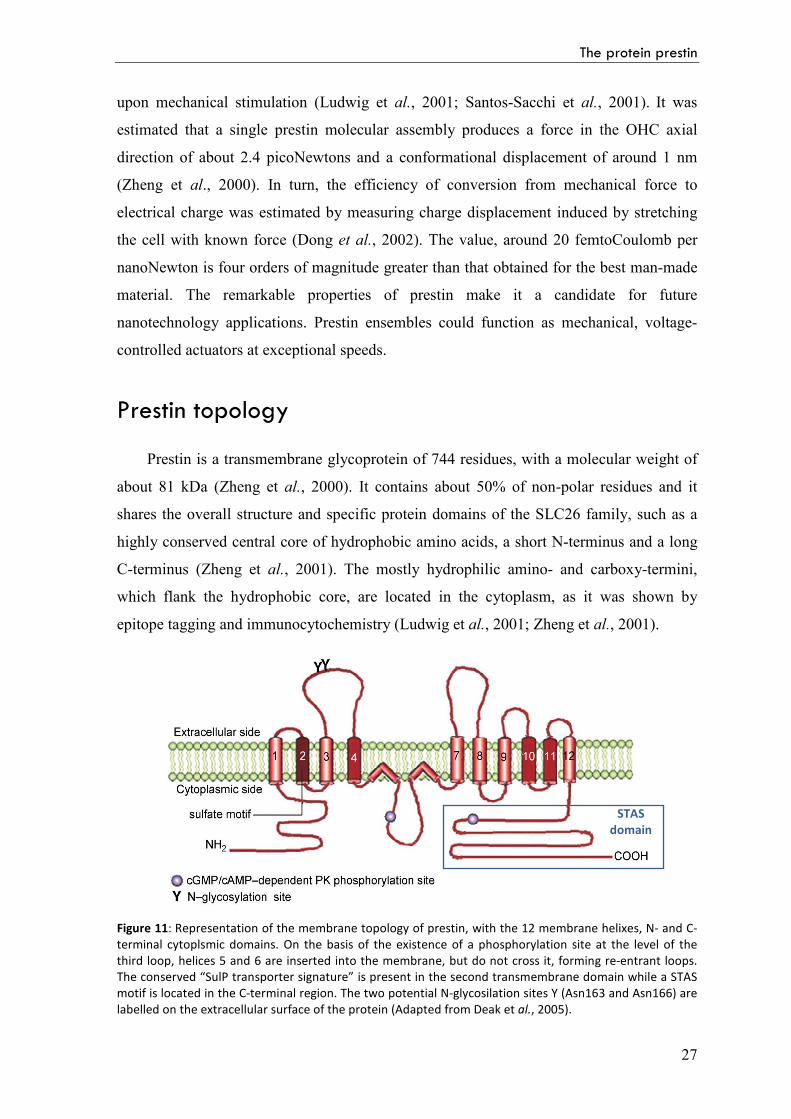
Prestin is a transmembrane glycoprotein of 744 residues, with a molecular weight of
about 81 kDa (Zheng et al., 2000). It contains about 50% of non-polar residues and it
shares the overall structure and specific protein domains of the SLC26 family, such as a
highly conserved central core of hydrophobic amino acids, a short N-terminus and a long
C-terminus (Zheng et al., 2001). The mostly hydrophilic amino- and carboxy-termini,
which flank the hydrophobic core, are located in the cytoplasm, as it was shown by
epitope tagging and immunocytochemistry (Ludwig et al., 2001; Zheng et al., 2001).
Figure 11: Representation of the membrane topology of prestin, with the 12 membrane helixes, N- and C-terminal cytoplsmic domains. On the basis of the existence of a phosphorylation site at the level of the third loop, helices 5 and 6 are inserted into the membrane, but do not cross it, forming re-entrant loops. The conserved “SulP transporter signature” is present in the second transmembrane domain while a STAS motif is located in the C-terminal region. The two potential N-glycosilation sites Y (Asn163 and Asn166) are labelled on the extracellular surface of the protein (Adapted from Deak et al., 2005).
STAS
domain
Part A: Introduction
28
The number of the membrane helixes is still disputed as topology prediction
programs produce ambiguous results: 10 or 12 transmembrane helixes can be
hypothesized (Oliver at al., 2001; Zheng at al., 2001; Deak et al., 2005; Navaratnam et al.,
2005;). The 12 transmembrane domains model is supported by more experimental
evidence and it is, in part, based on placing two potential N-glycosilation sites (Asn163
and Asn166) on the extracellular surface of the protein (Matsuda et al., 2004). In Figure
11, prestin is represented with 12 membrane helixes: on the basis of the existence of a
phosphorylation site (cGMP/cAMP-dependent PK phosporylation syte) at the level of the
third loop, helices 5 and 6 are inserted into the membrane, but do not cross it, forming re-
entrant loops (Deak et al., 2005).
The conserved “SulP transporter signature” is present in the second transmembrane
domain, while the C-terminal cytoplasmic region includes the Sulphate Transporter and
Anti-Sigma factor antagonist (STAS) domain. Two distinctive charged segments are
located in the C-terminal region: a positive-charge cluster is located at residues 557-580;
adjacent to this there is a negative-charge cluster at residues 596-613.
Although prestin is most closely related to SLC26A6, the human and mouse
orthologs of A6 have only 78% amino acid identity. In contrast, prestin is a highly
conserved protein with 92.7% of amino acids being identical among four different
mammalian species: human, mouse, rat and gerbil. Such a high degree of conservation is
not common among other SLC26A members. Significant changes in prestin primary
sequence occurred after the split between mammalian and avian lines, suggesting that
prestin evolved in order to fit special mammalian needs (Dallos et al., 2006).
Mechanism of action
Prestin is a new type of biological motor. It is entirely different from the conventional
enzymatic-activity-based motor proteins, in that it does not need ATP to function, but it is
a direct voltage to force converter. In this case the energy is supplied by the changing
membrane potential of the cell and this is probably unique in the animal kingdom (Dallos
et al., 2006). The action of prestin is also orders of magnitude faster than that of any other
cellular motor protein, as it functions at microsecond rates. In fact, OHC motility works at
frequencies up to at least 70 kHz (Frank et al., 1999).
Although prestin possesses all the sequence domains conserved throughout the
SLC26 family, it has not yet been shown to function as an anion transporter. Moreover,
The protein prestin
29
neither gating charge movements nor a NLC have been reported for any other member of
the SLC26 family, suggesting that prestin may have a unique function within the family.
How the membrane potential change of OHCs results in structural changes in prestin,
corresponding to the motor function, is not understood yet. Conceptually, prestin should
comprise at least two essential functional domains: the voltage sensor that detects changes
in the transmembrane potential of the cell, and the actuator that undergoes a
conformational change and thereby facilitates cell contraction or elongation in response to
depolarization and hyperpolarization, respectively (Dallos and Fakler, 2002).
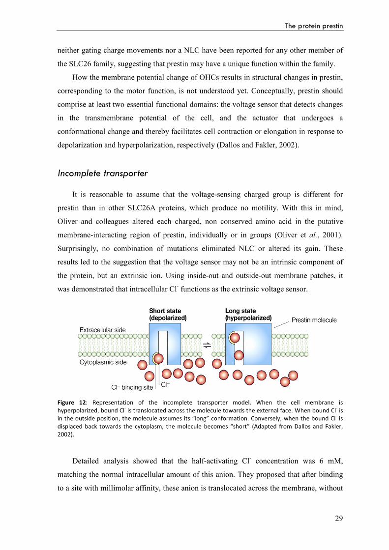
Incomplete transporter
It is reasonable to assume that the voltage-sensing charged group is different for
prestin than in other SLC26A proteins, which produce no motility. With this in mind,
Oliver and colleagues altered each charged, non conserved amino acid in the putative
membrane-interacting region of prestin, individually or in groups (Oliver et al., 2001).
Surprisingly, no combination of mutations eliminated NLC or altered its gain. These
results led to the suggestion that the voltage sensor may not be an intrinsic component of
the protein, but an extrinsic ion. Using inside-out and outside-out membrane patches, it
was demonstrated that intracellular Cl- functions as the extrinsic voltage sensor.
Figure 12: Representation of the incomplete transporter model. When the cell membrane is hyperpolarized, bound Cl- is translocated across the molecule towards the external face. When bound Cl- is in the outside position, the molecule assumes its “long” conformation. Conversely, when the bound Cl- is displaced back towards the cytoplasm, the molecule becomes “short” (Adapted from Dallos and Fakler, 2002).
Detailed analysis showed that the half-activating Cl- concentration was 6 mM,
matching the normal intracellular amount of this anion. They proposed that after binding
to a site with millimolar affinity, these anion is translocated across the membrane, without
Part A: Introduction
30
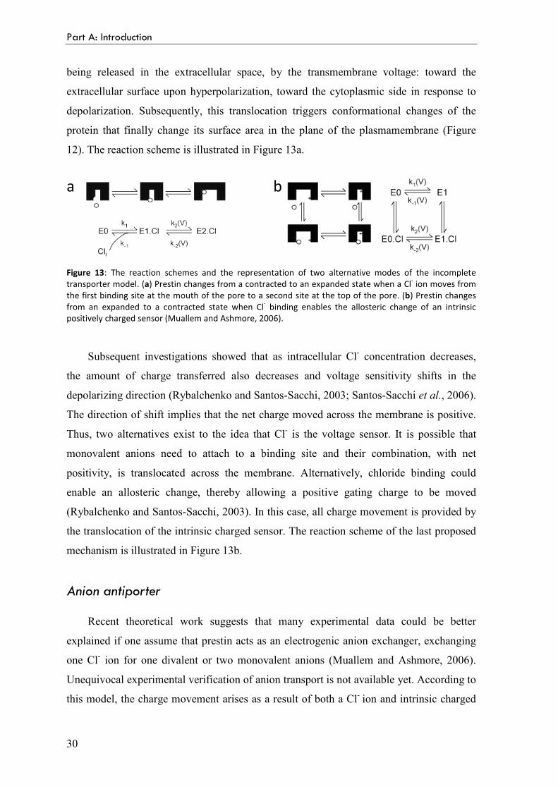
being released in the extracellular space, by the transmembrane voltage: toward the
extracellular surface upon hyperpolarization, toward the cytoplasmic side in response to
depolarization. Subsequently, this translocation triggers conformational changes of the
protein that finally change its surface area in the plane of the plasmamembrane (Figure
12). The reaction scheme is illustrated in Figure 13a.
Figure 13: The reaction schemes and the representation of two alternative modes of the incomplete transporter model. (a) Prestin changes from a contracted to an expanded state when a Cl- ion moves from the first binding site at the mouth of the pore to a second site at the top of the pore. (b) Prestin changes from an expanded to a contracted state when Cl- binding enables the allosteric change of an intrinsic positively charged sensor (Muallem and Ashmore, 2006).
Subsequent investigations showed that as intracellular Cl- concentration decreases,
the amount of charge transferred also decreases and voltage sensitivity shifts in the
depolarizing direction (Rybalchenko and Santos-Sacchi, 2003; Santos-Sacchi et al., 2006).
The direction of shift implies that the net charge moved across the membrane is positive.
Thus, two alternatives exist to the idea that Cl- is the voltage sensor. It is possible that
monovalent anions need to attach to a binding site and their combination, with net
positivity, is translocated across the membrane. Alternatively, chloride binding could
enable an allosteric change, thereby allowing a positive gating charge to be moved
(Rybalchenko and Santos-Sacchi, 2003). In this case, all charge movement is provided by
the translocation of the intrinsic charged sensor. The reaction scheme of the last proposed
mechanism is illustrated in Figure 13b.
Anion antiporter
Recent theoretical work suggests that many experimental data could be better
explained if one assume that prestin acts as an electrogenic anion exchanger, exchanging
one Cl- ion for one divalent or two monovalent anions (Muallem and Ashmore, 2006).
Unequivocal experimental verification of anion transport is not available yet. According to
this model, the charge movement arises as a result of both a Cl- ion and intrinsic charged
a b
The protein prestin
31
residues moving across the membrane. Thus net positive charge is moved across the
membrane as the Cl- ion is moved towards the extracellular surface. This model is
independent of the nature of the Cl- replacing anion which could be mono- or divalent as
long as it guarantees that the reorientation of the intrinsic charged residues is
electroneutral. The reaction scheme is illustrated in figure 14.
Figure 14: (a) The reaction scheme for a Cl-/SO42- exchanger model. Prestin exchanges one Cl- ion for one
SO42- ion via an alternating-access mechanism, in which prestin can only change between inward and
outward facing states with an anion bound. (b, c) Two alternative representations of the reaction scheme:
both assignments ensure that the critical voltage-dependent transition, E1.Cl↔E2.Cl, is associated with a conformational change of prestin into a compact state and symmetry is maintained (Muallem and Ashmore, 2006).
Prestin STAS domain
The intracellular C-terminus of prestin is the least well-conserved region compared
with other SLC26A proteins and it includes a STAS domain. It has only 25-35%
homology with its SLC26A relatives and it is expected to be responsible for the protein
specific function. Different experiments showed that changing charged amino acids in the
C-terminus to either the opposite charge (R, K > D; E, D > K) or a neutral amino acid (Q)
is not able to abolish NLC and does not disrupt plasma membrane (PM) targeting of
prestin (Oliver et al., 2001; Bai et al., 2006).
The role of the C-terminus of prestin was investigated in some detail by Dallos and
his group with a series of deletion, point and chimeric mutants (Zheng et al., 2005). The
function and cellular expression of mutants were examined in a heterologous expression
system (TSA-201 and OK cells) by measurement of NLC and confocal
immunofluorescence. The subcellular localization of mutant proteins was examined by co-
localization experiments of prestin with other subcellular component markers. The
a b c
Part A: Introduction
32
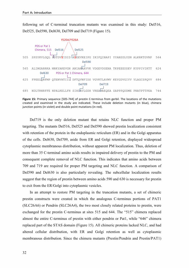
following set of C-terminal truncation mutants was examined in this study: Del516,
Del525, Del590, Del630, Del709 and Del719 (Figure 15).
Figure 15: Primary sequence [505-744] of prestin C-terminus from gerbil. The locations of the mutations created and examined in the study are indicated. These include deletion mutants (in blue), chimera junction points (in violet) and double point mutations (in red).
Del719 is the only deletion mutant that retains NLC function and proper PM
targeting. The mutants Del516, Del525 and Del590 showed prestin localization consistent
with retention of the protein in the endoplasmic reticulum (ER) and in the Golgi apparatus
of the cells. Del630, Del709, aside from ER and Golgi retention, displayed widespread
cytoplasmic membranous distribution, without apparent PM localization. Thus, deletion of
more than 35 C-terminal amino acids results in impaired delivery of prestin to the PM and
consequent complete removal of NLC function. This indicates that amino acids between
709 and 719 are required for proper PM targeting and NLC function. A comparison of
Del590 and Del630 is also particularly revealing. The subcellular localization results
suggest that the region of prestin between amino acids 590 and 630 is necessary for prestin
to exit from the ER/Golgi into cytoplasmic vesicles.
In an attempt to restore PM targeting in the truncation mutants, a set of chimeric
prestin constructs were created in which the analogous C-terminus portions of PAT1
(SLC26A6) or Pendrin (SLC26A4), the two most closely related proteins to prestin, were
exchanged for the prestin C-terminus at sites 515 and 644. The “515” chimera replaced
almost the entire C-terminus of prestin with either pendrin or Pat1, while “646” chimera
replaced part of the STAS domain (Figure 15). All chimeric proteins lacked NLC, and had
altered cellular distribution, with ER and Golgi retention as well as cytoplasmic
membranous distribution. Since the chimera mutants (Prestin/Pendrin and Prestin/PAT1)
The protein prestin
33
could not restore prestin PM targeting, the capacity for prestin to insert into the PM of
cultured epithelial cells may be dependent on prestin specific C-terminal amino acid
residues.
A tyrosine-containing motif (YXXΦ) is one of several well studied motifs that direct
the transport of newly synthesized membrane protein from the trans-Golgi network to the
basolateral membrane (Keller and Simons, 1997). In this motif Y is tyrosine, X is any
amino acid, and Φ is a bulky hydrophobic amino acid. There are seven potential tyrosine
containing motifs in the C-terminus of prestin. The aberrant PM targeting seen with the
deletion mutants and chimeric proteins may be related to the loss of potential basolateral
membrane targeting motifs located in the C-terminus. To approach the question whether
these motifs might be involved in membrane targeting of prestin to its PM location, a
double point mutant was created: Y520A/Y526A, which abolished two of the seven
potential tyrosine-containing motifs (Figure 15). The mutation Y520A/Y526A resulted in
lost of NLC function and in intracellular accumulation of prestin. These observations
indicate that specific sequences within the C-terminus are essential for the NLC function
in addition to its role in membrane targeting.
Altogether these data indicate that the C-terminus of prestin is likely to be intimately
involved in anion binding, membrane targeting, and the voltage regulated conformational
change of prestin. How the distal amino acids of the C-terminus regulate PM targeting and
protein function is not understood yet.
Oligomerization properties
The examination of OHCs membranes by freeze fracture reveals densely packed 11
nm diameter particles (Figure 9; Forge et al., 1991; Kalinec et al., 1992). It has been a
consistent assumption that the particles consist of some multimer of the motor protein,
inasmuch as the 744 amino acid prestin molecule is too small to produce an 11 nm
monomer. How prestin forms oligomers and what part of the molecule is involved in their
formation is not completely clear yet, although the involvement of both the N- and the C-
terminal domains has been suggested (Navaratnam et al., 2005; Zheng et al., 2005).
The first evidence for prestin multimerization came from fluorescence resonance
energy transfer experiments, that showed that homodimerization of prestin depends on an
intact N-terminus (Navaratnam et al., 2005; Greeson et al., 2006).
Part A: Introduction
34
The number of subunits necessary to form a functional motor protein was first
addressed by Zheng and colleagues (Zheng et al., 2006). In this study, native and
recombinant prestin, obtained from different expression systems, including yeast and
mammalian cell lines, was seen resistant to dissociation by lithium dodecyl sulphate
(LDS) and behaving as a stable oligomer on LDS-PAGE. Chemical cross-linking and
perfluoro-octanoate-electrophoresis (PFO-PAGE) combined with immunoblotting and
affinity purification suggest a tetrameric subunit stoichiometry of prestin. Moreover
sodium dodecyl sulphate (SDS) dissociates the tetramer into dimers that could be
converted to monomers by hydrophobic reducing agents, but not by the hydrophilic ones.
These data suggest that prestin monomers are covalently linked to dimers by disulfide
bonds located in the hydrophobic membrane core and that these covalently linked dimers
associate via hydrophobic interactions to form a tetramer. They proposed that the stable
covalent dimer may act as the building block for producing the higher order oligomers that
form the 11 nm particles in the OHC basolateral membrane.
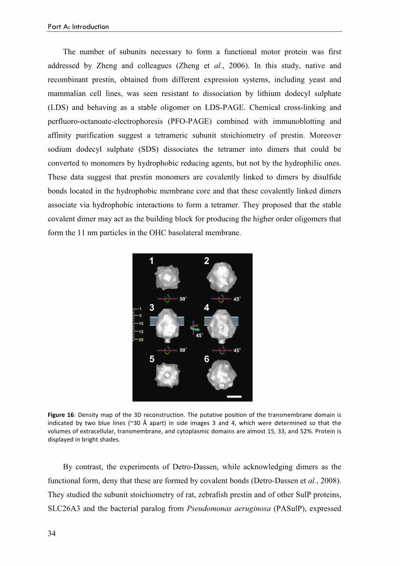
Figure 16: Density map of the 3D reconstruction. The putative position of the transmembrane domain is indicated by two blue lines (~30 Å apart) in side images 3 and 4, which were determined so that the volumes of extracellular, transmembrane, and cytoplasmic domains are almost 15, 33, and 52%. Protein is displayed in bright shades.
By contrast, the experiments of Detro-Dassen, while acknowledging dimers as the
functional form, deny that these are formed by covalent bonds (Detro-Dassen et al., 2008).
They studied the subunit stoichiometry of rat, zebrafish prestin and of other SulP proteins,
SLC26A3 and the bacterial paralog from Pseudomonas aeruginosa (PASulP), expressed
The protein prestin
35
in Xenopus laevis oocytes or in mammalian cells. According to blue native PAGE and
chemical cross-linking experiments, prestin and the other SulP proteins form dimers as
predominant oligomeric state. Oligomers dissociate entirely into monomers under non-
reducing conditions in the presence of low concentrations of SDS. So they concluded that
dimers are held together by non-covalent forces rather than by covalent disulfide bonds.
A preliminary indication of prestin shape was provided by Mio and colleagues who
expressed prestin in baculovirus-infected Sf9 cells and purified it (Mio et al., 2008). They
observed the negatively stained molecules using electron microscopy, and reconstructed
the 3D structure of prestin at 2 nm resolution by single particle analysis. Their result is
consistent with prestin being a tetramer, having a large cytoplasmic domain and assuming
a “bullet shape”, with a fourfold symmetry (Figure 16).
Prestin orthologs
A recent development in the study of prestin is the analysis of its orthologs. Indeed,
when assessed by sequence similarity, the closest homolog of mammalian SLC26A5 is
that found in the zebrafish hearing organ (Albert et al., 2007). The zebrafish prestin
orthologue, zprestin, shares around 50% amino acid identity with mammalian prestin.
Like its mammalian orthologue, zprestin is expressed in hair cells of the ear and confers
NLC to the membranes of transfected cells, similar to the characteristic electrogenic
charge movement that accompanies the prestin-mediated somatic electromotility of
mammalian OHCs. Although expression analysis and electrophysiological data show that
zprestin properly localizes to the cell membrane upon heterologous expression, it
nonetheless fails to generate electromotile responses of the transfected cells. Hence,
though displaying a prestin-like voltage sensitivity, zprestin does not seem to be a prestin-
like motor, supporting the general view that a prestin-mediated somatic electromotility is a
unique feature of mammalian OHCs.
The transport function of zprestin was tested, revealing that it is an electrogenic
divalent/chloride anion antiporter, exchanging sulphate or oxalate for chloride in a strictly
coupled manner with a 1:1 stoichiometry (Schaechinger and Oliver, 2007). The same
result was obtained with the chicken ortholog, which most probably lacks electromotility.
Thus, the prestin orthologs from chicken and zebrafish appear to be an intermediate
form, performing voltage-dependent, chloride-sensitive charge dislocations, including full
anion transport, but not having acquired motility. The presence of prestin transporter in
Part A: Introduction
36
non-mammalian hair cells makes sense in evolutionary terms: a protein already present in
the hair cells of phylogenetic ancestors to mammals may have adopted a novel,
electromotile function during evolution toward the mammalian outer hair cell. Zebrafish
and chicken prestin can thus be regarded as the “missing link” that may help us to
understand the sequence of events that has led to the emergence of a novel type of motor
protein in the course of SLC26 evolution.
22
TThhee pprroojjeecctt
The project
39
Aims of this study
Despite the increasing interest in the SLC26 genes and, in general, in the SulP
members, a substantial amount of research is still needed to understand the roles of these
transporters. In particular, very little is known about the structural organization of these
proteins and no three-dimensional structure of domains or full-length sequences are
available for any mammalian SLC26 anion transporter or for other members of the SulP
family, of any species. The structural characterization is fundamental for the
comprehension of the mode of action of a protein and it is an essential step for the
understanding of the functional consequences of the mutations responsible for related
pathologies. In this context, the long term task is the elaboration of a molecular functional
model of the SulP anion transporters, able to explain both the common features of this
class of transporters and the peculiar characteristics of the single components.
To this purpose, first important step is the structural characterization of the
functionally important C-terminal STAS domain. The work described in this thesis has
been focused on the production of different forms of the STAS domain, for the
biophysical and structural characterization. The second part of the project is more
ambitious and concerns the production of the full-length membrane proteins by a cell-free
expression system.
The strategy
To characterize a protein from a structural point of view, it is necessary to produce it
in amounts in the order of milligrams. Due to the low abundance of the SulP transporters
in natural sources, the unavoidable choice is the production of recombinant material. The
strategy adopted includes amplification of the selected genes, starting from the cDNA of
the related proteins, cloning into appropriate bacterial expression plasmids, expression in
E. coli and purification. All the used expression vectors produce a recombinant protein
linked to another protein or a short peptide with well-known properties (the so-called
“tags”). These fusion tags help to isolate the protein of interest by affinity
chromatography. Subsequently, the recombinant protein is excided from the tag by an
appropriate proteolytic enzyme and further purified. For the structural and biophysical
characterization, several complementary techniques are used, such as circular dichroism
Part A
40
(CD) and fluorescence spectroscopy, dynamic light scattering (DLS), and, if possible, X-
ray crystallography, for the characterization at atomic level.
Since the size of the STAS domain is suitable, it is characterized also by solution
NMR, in collaboration with the group of Prof. Stefano Mammi from the Department of
Chemical Sciences of the University of Padova. The joint crystallographic and NMR
efforts may provide a complete structural characterization of the STAS domains, giving
complementary information. Crystallography can reveal the high resolution structural
details of the STAS domains and their binding properties to tightly bound small molecules
and ions. NMR can complete the structural characterization by providing information
about the flexibility and dynamics of the domain in solution (most of the variable loop is
predicted disordered), and the binding properties to medium or low affinity species.
For the production of the full-length membrane proteins, a variety of intrinsic
problems exists with the commonly employed heterologous expression systems. Cell-free
expression system represents a recently developed and powerful alternative to in vivo
expression.
Production and characterization of the STAS domain
The STAS domains of different SulP proteins, from distance-related species, are
selected for the structural and biophysical characterization: prestin (SLC26A5) from
Rattus norvegicus and Meriones unguiculatus, pendrin (SLC26A4) from Homo sapiens
and the bacterial sulphate transporter Rv1739c from Mycobacterium tuberculosis.
The attention is focused on mammalian pendrin and prestin, because they share
interesting properties: both are expressed in the inner ear and mutations in each of the two
genes are linked to deafness or hearing impairment. Pendrin is an extremely interesting
target also because the Pendred syndrome is genetically well characterized.
The study of mycobacterial SulP transporters offers potential insight into the sulphur
assimilation pathways leading to biosynthesis of sulfolipid pathogenicity determinants.
Moreover, the structural characterization of Rv1739c STAS domain may shed light on the
evolution mechanisms of this domain and in the different roles played in the various
transporters. This approach can be also “technically” useful, since it is well known that
even similar polypeptides can have substantially different properties in terms of
propensities to crystallize or NMR feasibility.
The project
41
In order to identify a sequence corresponding to a compact single domain, several
types of analyses and predictions are performed on the C-terminal part of each SulP
protein, such as multiple sequence analyses, secondary structure predictions, predictions
of intrinsic disordered regions and homology modeling outputs. The accurate selection of
the N- and C-termini is more critical for domains that are part of a larger protein, as it is
the case of the SulP STAS domains, whose boundaries are not clearly defined by sequence
alignments. For this reason constructs of different length are selected, for each SulP
protein. In parallel, information from literature, in particular functional data on mutations
and deletions, are taken into account.
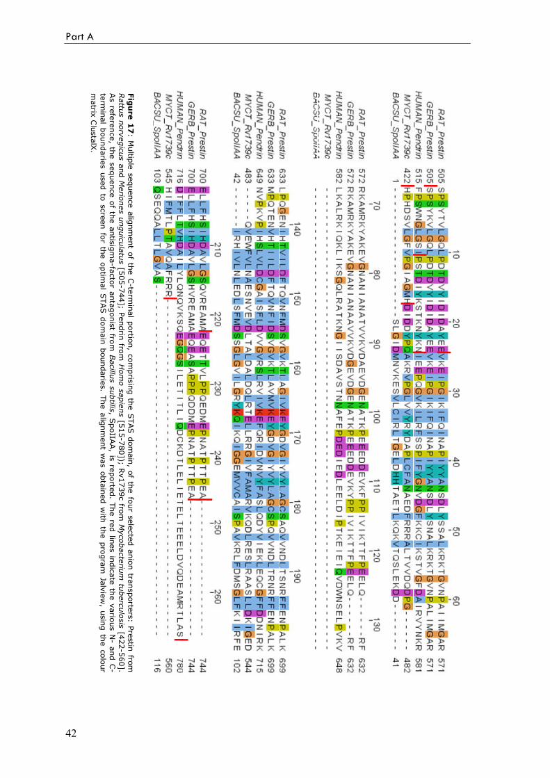
From the sequence alignment in Figure 17 it is immediately evident that the sequence
similarity between mammalian transporters and Rv1739c C-terminal region is very low,
(less than 20%). Most of the difference is found in the long insertion (70, 80 ammino acids
long, between the position 60 and 140 in the alignment) and in the longer C-terminal
extension present in the mammalian transporters STAS domain. The evolutionary and
functional role of the insertion is unknown; secondary structure predictions suggest it is
largely unstructured. According to these considerations, Rv179c STAS domain is more
similar to the ASA protein STAS domain (SpoIIAA from Bacillus subtilis is reported in
the alignment as reference). Taking into account these differences in lengths as well as the
low amino acid conservation observed, most probably the 3D structure of SulP STAS is
significantly different from that of the ASA proteins. Presumably, the differences are
responsible for the distinct properties of this domain when part of the different
transporters.
Part A
42
Figure 17: M
ultip
le sequence alignment o
f the C-te
rminal p
ortio
n, c
omprising th
e STAS domain, o
f the fo
ur s
elected anion tra
nsporte
rs: P
restin
from
Rattus norvegicus and Meriones unguiculatus [5
05-744]; P
endrin
from Homo sapiens [5
15-780]); R
v1739c fro
m Mycobacterium tuberculosis [4
22-560].
As re
ference, th
e sequence of th
e antisigma-fa
ctor a
ntagonist fro
m Bacillus subtilis
, SpoIIA
A, is
reporte
d. T
he re
d lin
es in
dicate th
e vario
us N- a
nd C-
term
inal boundarie
s used to screen for the optim
al STAS domain boundarie
s. The alignment was obtained with
the program Jalview, using the colour
matrix
ClustalX.
The project
43
Production of SulP proteins by cell-free expression system
The structural characterization of membrane proteins is still a challenge mainly
because of the difficulty to produce adequate amount (in the order of milligrams) of
purified and functional material (Lacapere et al., 2007). This is particularly true for
eukaryotic membrane proteins, whose high resolution 3D structure is known only for a
very few of them.
The production of full-length prestin is first tested for over-expression in classical
bacterial systems. The prestin gene is cloned into an expression vector (pET151/D-TOPO,
Invitrogen) that allows the production of a recombinant fusion protein with a His-tag at
the N-terminus. Different E. coli strains and different expression conditions are tested, in
particular E. coli strains C41(DE3) and C43(DE3) (Miroux and Walker, 1996) that were
reported successful in the expression of other membrane proteins (Dumon-Seignovert et
al., 2004).
As alternative strategy, the protein is produced with cell-free systems based on E. coli
extracts, an emerging and really promising technique for the large-scale production of
membrane proteins. As for the STAS domain, different SulP proteins are selected for cell-
free expression: prestin (SLC26A5) from rat, zebrafish and chicken; the transporter
Rv1739c from Mycobacterium tuberculosis; the plant transporter SULTR1.2 from
Arabidopsis thaliana.
33
RReessuullttss
aanndd
ddiissccuussssiioonn
47
3.1 Overview
Three variants of prestin C-terminal domain were cloned and produced: one from
mongolian gerbil and two from rat. Two constructs were selected either for human pendrin
or for the bacterial protein, Rv1739c. (Figure 17). The Table 2 summarizes the cloning
and expression results for each of the selected constructs (for a total of 7 constructs).
Table 2: Survey of the STAS domains cloned and expressed in E. coli.
STAS
domain Species Sequence AA
MW
(kDa)
Expression
vector Tag protein
Expression/
Solubility
PreCDT M. unguiculatus [505-744] 240 26.6 pET-19 (His)10 Low/ Good
PreCDL R. norvegicus [529-744] 216 23.8 pGEX-4T
pET-28
GST
(His)6
No detectable
Good/ High
PreCDS R. norvegicus [529-720] 192 21.2 pGEX-4T
pET-28
GST
(His)6
No detectable
Good/ Good
PenCDL H. sapiens [525-780] 256 29.1 pET-151 (His)6 Medium/ Low
PenCDS H. sapiens [539-780] 242 27.5 pET-151 (His)6 Medium/ Low
RvCDL M. tuberculosis [422-560] 139 15.7 pET-SUMO (His)6-SUMO High/ Good
RvCDS M. tuberculosis [439-560] 122 14.0 pET-SUMO (His)6-SUMO High/ Good
Refmac) with visual inspection of the electron density map and manual adjustment by
using the program Coot (Emsley and Cowtan, 2004). During refinement, water molecules
were added to the model, both automatically and manually, and those with B factors
higher than 50 were excluded. Some molecules of MPD were also introduced and refined.
Figure 6: Crystals of GFPmut2.
Structure of the GFPmut2
113
Table 1: Data collection and refinement statistics of GFPmut2 at both pH 6 and 9.
GFPmut2 pH 6.0 pH 9.0
Data collection (XRD1, ELETTRA, Trieste)
Wavelength(Å) 1.1 1.2
Space Group P 212121 P 212121
Axes a, b, c (Å) 32.5 59.9 112.4 32.5 59.9 112.1
Resolution (Å) 1.64 (1.72) 1.66 (1.75)
Independent reflections 23039 (2269) 21006 (2204)
Multiplicity 4.0 (3.4) 5.7 (5.6)
Completeness (%) 82.9 (82.9) 78.6 (78.6)
Rsym 0.079 (0.270) 0.061 (0.241)
Rmeas 0.090 (0.316) 0.068 (0.265)
<I/σ (I)> 7.3 (2.1) 8.9 (3.0)
BWilson (Å2) 11.4 15.1
Refinement
Protein Residues 233 233
Solvent molecules 287 234
Other molecules 2 MPD 3 MPD
Rwork/Rfreea (%) 17.2/ 21.1 16.9/ 21.1
Bmean (Å2) 10.84 9.73
Numbers in parentheses refer to the last resolution shell. a R free was calculated using 5% of the data.
Result and discussion
Crystal structures
The crystal structure of GFPmut 2 at both pH 6 and pH 9 (above and below the pKa
in the crystal) was solved to 1.64 and 1.66 Å resolution, respectively, in order to
investigate the structural basis of the spectral response to changes in pH. The overall
structure is very similar to that of the wtGFP: 11 β-strands form the classic “β-can”
structure and the chromophore, belonging to the central distorted helix, is well buried
inside (Figure 7). The quality of the models is good (Figure 8) and the high resolution
allows a detailed analysis of the chromophore environment.
Part B: Results and discussion
114
Figure 7: Ribbon view of GFPmut2, seen from the front and from above. The β-sheets are displayed in yellow, the helixes in red and the loops in green. The chromophore is shown as a ball-and-stick model. The figures were produced by PyMol.
His148
Thr203
Glu222
Tyr66
Ala65
Gly67
Figure 8: Stereo image of (2Fo-Fc) electron density map, contoured at 1.0 σ, and atomic model of GFPmut2 at pH 6, showing the fluorophore and its environment at around 1.6 Å resolution.
Structure of the GFPmut2
115
The main differences compared to both the wtGFP (PDB code 1GFL; Yang et al.,
1996) and the S65T mutant (PDB code: 1EMG; Elsliger et al., 1999) are found in the
chromophore position and in the surrounding water molecules and can be ascribed to
mutations proper to GFPmut2, in particular Ser65Ala and Val68Leu. Due to the bulkier
side chain compared to valine, leucine 68 causes a slight shift of the chromophore toward
the β-sheet external wall of the protein, in particular toward His148 now at only 3.2 Å in
the pH 6.0 structure (with the protonated chromophore). This allows a direct interaction
between the two residues (Figure 9), with the formation of a hydrogen bond not possible
in other GFP structures with the chromophore in the protonated form, such as the wtGFP
(PDB code 1GFL, Figure 11).
His148
Leu68
Ser205 Glu222
Chromophore
Ala65
W2
W3 W5
W4
W1
Thr203
Figure 9: Stereo image of the atomic model of the fluorophore and its environment in the GFPmut2 mutant at pH 6. Ala65, Leu68, His148, Thr203, Ser205, Glu222 and water molecules (W1, W2, W3, W4, W5) are labelled. Dashed lines indicate hydrogen bonds.
Another interesting feature of the GFPmut2 structures is the presence of two
additional water molecules (W4 and W5 in Figure 9) in the proximity of the chromophore,
in the plane roughly formed by Glu222, Gln69 and Thr203. Usually, here two water
molecules (W2 and W3 in Figures 11, 12) are found, connecting Glu222 and Gln69.
Thr203 side chain usually interacts directly with only one water molecule (or none in the
S65T mutant). In GFPmut2, the position of the two waters (W2 and W3) is different and
Part B: Results and discussion
116
two additional water molecules are found (W4 and W5), with the consequences that
Glu222 and Thr203 are now directly bridged by W2 and W3 and that Thr203 side chain
conformation is stabilized by the interactions with W3 and W5, at 2.6 and 2.7 Å. This is
quite relevant because Thr203 side chain usually rotates away from this position in the
presence of a deprotonated chromophore (such as in the structure of S65T mutant at pH 8,
Figure 12), contributing to its stabilization with a direct H-bond. In GFPmut2 this
movement is much more difficult as the Thr hydroxyl group is “fixed” by the two
hydrogen bonds with W3 and W5. Another important characteristic of the mut2 structures
is that the lack of a hydroxyl function in residue 65 (Ser in wt, Figure 11; Thr in S65T
mutant, Figure 12) hampers the possibility to Glu222 to interact with such function.
Therefore, Glu222 always interacts with Ser205, taking part in a sort of “circular” H-bond
network that now extends from the chromophore hydroxyl to Thr203, through W1,
Ser205, Glu222 and two other water molecules,W2 and W3 (Figure 9).
In the structure determined at pH 9 there is only one relevant change, the distance
between the hydroxyl chromophore and His148 becomes smaller, from 3.20 to 2.92 Å,
indicating a stronger interaction between the two, probably for the deprotonation of the
chromophore that donates the proton to the His imidazole ring in an acid-base exchange
illustrated in Figure 10. This is well in accordance with the spectroscopic studies of the
GFPmut2 properties in solution, which revealed how His148 acts as the primary acceptor
for protons coming from the bulk, and their following release to the phenolic oxyanion of
the chromophore with relatively high efficiency (Abbruzzetti et al., 2005).
At variance with the wt and other GFP mutants studied so far, in mut2 the
chromophore deprotonation is not coupled with conformational rearrangements of the
surrounding residues, with particular reference to Thr203 and Glu222, now more strongly
anchored in their position by the more extended H-bonding network.
The difference between the pKa calculated in solution (around 6) and that in the
crystal (around 7) is probably due to the presence of the high MPD concentration in the
crystallization medium.
OHN N
H
Tyr66
ON N H
Tyr66
OH
H+
His148 His148
Figure 10: Acid-base equilibrium between His148 and Tyr66.
Structure of the GFPmut2
117
His148
Val68
Ser205Glu222
Ser65W3
W2 W1
Chromophore
Thr203
Figure 11: Stereo image of the atomic model of the fluorophore in the protonated form and its environment in the wtGFP (PDB code 1GFL; Yang et al., 1996). Ser65, Val68, His148, Thr203, Ser205, Glu222 and water molecules (W1, W2, W3) are labelled. Dashed lines indicate hydrogen bonds.
His148
Ser205 Glu222
Thr65
Val68
W3
W2
W1
Chromophore
Thr203
Figure 12: Stereo image of the atomic model of the fluorophore in the deprotonated form and its environment in the S65T mutant at pH 8 (PDB code 1EMG; Elsliger et al., 1999). Thr65, Val68, His148, Thr203, Ser205, Glu222 and water molecules (W1, W2, W3) are labelled. Dashed lines indicate hydrogen bonds.
Part B: Results and discussion
118
We can conclude that the crystal structures of GFPmut2 at two different pH have
indeed revealed the structural bases of the spectral properties of this mutant. The
mutations, in particular Ser65Ala and Val68Leu, are responsible for an intrinsically closer
interaction between the chromophore and His148. Moreover, the presence of a more
extended H-bonding network involving Thr203 and Glu222 is responsible for their
diminished conformational freedom.
119
RReeffeerreenncceess
Abbruzzetti S., Grandi E., Viappiani C., Bologna S., Campanini B., Raboni S., Bettati S.,
Mozzarelli A. (2005). Kinetics of Acid-Induced Spectral Changes in the GFPmut2
Chromophore. J. Am. Chem. Soc. 127: 626-635.
Bizzarri R., Serresi M., Luin S., Beltram F. (2008). Green fluorescent protein based pH
indicators for in vivo use: a review. Anal. Bioanal. Chem. DOI: 10.1007/s00216-008-
2515-9.
Brejc K., Sixma T.K., Kitts P.A., Kain S.R., Tsien R.Y., Ormo M., Remington S.J. (1997).
Structural basis for dual excitation and photoisomerization of the Aequorea victoria green
fluorescent protein. Proc. �atl. Acad. Sci. USA 94: 2306-2311.
Chalfie M., Tu Y., Euskirchen G., Ward W.W., Prasher D.C. (1994). Green fluorescent
protein as a marker for gene expression. Science 263: 802-805.
Chirico G., Cannone F., Beretta S., Diaspro A., Campanini B., Bettati S., Ruotolo R.,
Mozzarelli A. (2002). Dynamics of green fluorescent protein mutant2 in solution, on spin-
coated glasses, and encapsulated in wet silica gels. Protein Sci. 11: 1152-1161.
Chudakov D.M., Lukyanov S., Lukyanov K.A. (2005). Fluorescent proteins as a toolkit
for in vivo imaging. Trends Biotechnol. 23: 605-613.
Collaborative Computational Project N. 4. The CCP4 suite: programs for protein
crystallography. (1994). Acta Crystallogr. D 50: 760-763.
Cormack B.P., Valdivia R.H., Falkow S. (1996). FACS-optimized mutants of the green
fluorescent protein (GFP). Gene 173: 33-38.
Part B
120
Cubitt A.B., Heim R., Adams S.R., Boyd A.E., Gross L.A., Tsien R.Y. (1995).
Understanding, improving and using green fluorescent proteins. Trends Biochem. Sci. 20: