

““RIABILITAZIONE DELLE RIABILITAZIONE DELLE MALATTIE MALATTIE
NEUROMUSCOLARINEUROMUSCOLARI””
Neuromuscular DisordersNeuromuscular Disorders

Prevalenceper 100.000Most Common
Prevalenceper 100.000LessCommon
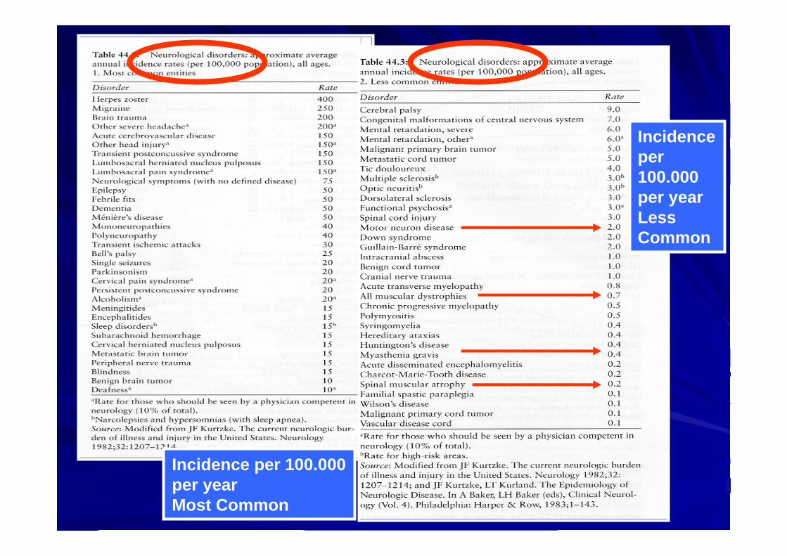
Incidence per 100.000per yearMost Common
Incidenceper 100.000 per yearLessCommon

NEUROMUSCULAR DISEASES NEUROMUSCULAR DISEASES ((acquiredacquired or or hereditaryhereditary))
- Muscle- Neuromuscular
junction- Nerve
- Nerve plusCentralNervousSystem
C. Current concept: individual muscle fibersof same motor unit are scattered throughoutmuscle

Neuromuscular disorders: Neuromuscular disorders: –– Muscle weaknessMuscle weakness::
Amyotrophic lateral sclerosis ALS (Lou Amyotrophic lateral sclerosis ALS (Lou GerigGerig’’ssDisease)Disease)CharcotCharcot--MarieMarie--Tooth syndromeTooth syndromeMyasthenia gravis Myasthenia gravis Muscular DystrophyMuscular Dystrophy
–– Movement disordersMovement disordersParkinsonParkinson’’s Diseases Disease
–– Combined muscle weakness and movement Combined muscle weakness and movement disorderdisorder
Multiple sclerosisMultiple sclerosis

Amyotrophic lateral sclerosis Amyotrophic lateral sclerosis ((ALSALS-- Lou GehrigLou Gehrig’’s disease)s disease)
Is a degenerative disease that affects the Is a degenerative disease that affects the upper and/or lower upper and/or lower motor neuronsmotor neurons (nerve cells controlling muscles) in the anterior (nerve cells controlling muscles) in the anterior (motor) horns of the spinal cord, and/or motor nuclei of the (motor) horns of the spinal cord, and/or motor nuclei of the lower brain.lower brain.
A disease of progressive loss of motor nerve cells in the brain A disease of progressive loss of motor nerve cells in the brain and spinal cord, causing progressive loss of motor controland spinal cord, causing progressive loss of motor control
As nerves die, the muscles atrophy. As nerves die, the muscles atrophy.

Symptoms of ALSSymptoms of ALSSymptoms usually do not develop until until after age 50, Symptoms usually do not develop until until after age 50, usually 5th or 6th decade of lifeusually 5th or 6th decade of life
Affects males more than femalesAffects males more than females
Symptoms begin with Symptoms begin with muscle weaknessmuscle weakness, , muscle crampsmuscle cramps, , and decrease in muscle strength and coordination that and decrease in muscle strength and coordination that eventually lead to eventually lead to paralysisparalysis..
There may be There may be muscle tremorsmuscle tremors, , spasmsspasms, twitching, or , twitching, or muscle atrophymuscle atrophy. .
Reflexes may be abnormal, Reflexes may be abnormal, including loss of the gag including loss of the gag reflexreflex. . Some patients have "emotional incontinence"Some patients have "emotional incontinence"
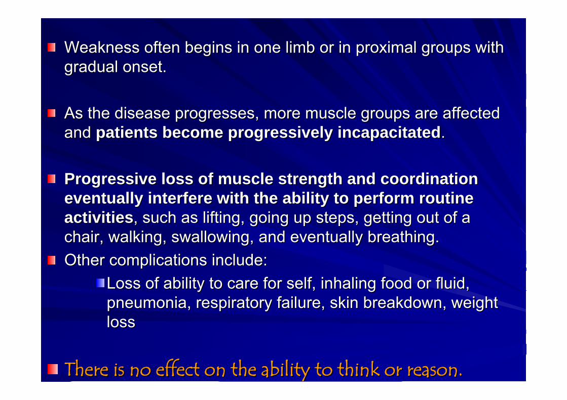
Weakness often begins in one limb or in Weakness often begins in one limb or in proximalproximal groups with groups with gradual onset. gradual onset.
As the disease progresses, more muscle groups are affected As the disease progresses, more muscle groups are affected and and patients become progressively incapacitatedpatients become progressively incapacitated. .
Progressive loss of muscle strength and coordination Progressive loss of muscle strength and coordination eventually interfere with the ability to perform routine eventually interfere with the ability to perform routine activitiesactivities, such as lifting, going up steps, getting out of a , such as lifting, going up steps, getting out of a chair, walking, swallowing, and eventually breathing. chair, walking, swallowing, and eventually breathing. Other complications include:Other complications include:
Loss of ability to care for self, inhaling food or fluid, Loss of ability to care for self, inhaling food or fluid, pneumoniapneumonia, respiratory failure, skin breakdown, weight , respiratory failure, skin breakdown, weight loss loss
There is no effect on the ability to think or reason.There is no effect on the ability to think or reason.
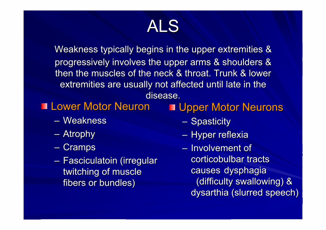
ALSALSWeakness typically begins in the upper extremities &Weakness typically begins in the upper extremities &progressively involves the upper arms & shoulders & progressively involves the upper arms & shoulders & then the muscles of the neck & throat. Trunk & lower then the muscles of the neck & throat. Trunk & lower extremities are usually not affected until late in the extremities are usually not affected until late in the
disease.disease.Lower Motor NeuronLower Motor Neuron–– WeaknessWeakness–– AtrophyAtrophy–– CrampsCramps–– FasciculatoinFasciculatoin (irregular (irregular
twitching of muscle twitching of muscle fibers or bundles)fibers or bundles)
Upper Motor NeuronsUpper Motor Neurons–– Spasticity Spasticity –– Hyper Hyper reflexiareflexia–– Involvement of Involvement of
corticobulbarcorticobulbar tracts tracts causes causes dysphagiadysphagia
(difficulty swallowing) & (difficulty swallowing) & dysarthiadysarthia (slurred speech)(slurred speech)

ALSALS——Lou GehrigLou Gehrig’’s diseases diseaseSigns and SymptomsSigns and Symptoms
Fatigue Fatigue Difficult doing motor task Difficult doing motor task –– Buttoning a shirtButtoning a shirt
Progressive muscle Progressive muscle weakness&wasting,atrophyweakness&wasting,atrophy–– When When intercostalintercostal muscles muscles
& diaphragm become & diaphragm become involved, involved, respresp are shallow are shallow & coughing is ineffective.& coughing is ineffective.
DysphagiaDysphagia--swallowingswallowingDysarthriaDysarthria --speechspeech–– Weakness begins in brain Weakness begins in brain
stem causing problems stem causing problems with speech & swallowing with speech & swallowing = bulbar ALS= bulbar ALS
Wt lossWt lossTongue fasciculationTongue fasciculation--twitchingtwitchingJaw Jaw clonusclonus ----involuntary involuntary tightening/ relaxing of tightening/ relaxing of musclesmusclesSpasticity of flexor musclesSpasticity of flexor muscles
Respiratory difficultyRespiratory difficulty--death occurs 2dary to death occurs 2dary to respresp infectioninfection

ALSALS——Lou GehrigLou Gehrig’’s diseases diseaseS/S continuedS/S continued
Involvement of upper an Involvement of upper an lower extremitieslower extremities
One side more than otherOne side more than other
No sensory lossNo sensory loss
Patient remains alertPatient remains alert
Death occurs 5 to 10 years Death occurs 5 to 10 years post diagnosispost diagnosis
Death usually results Death usually results from respiratory infectionfrom respiratory infectionsecondary to compromised secondary to compromised respiratory function.Caused respiratory function.Caused by respiratory or bulbar by respiratory or bulbar paralysisparalysis

Chief SymptomsChief Symptoms of ALSof ALSProgressive muscle weaknessProgressive muscle weakness
Atrophy/Atrophy/fasiculationsfasiculations (tongue twitching)(tongue twitching)
Spasticity with brisk overactive stretch reflexSpasticity with brisk overactive stretch reflex
Anal/bladder muscle weaknessAnal/bladder muscle weakness
With cranial nerves, resulting With cranial nerves, resulting dyarthriasdyarthrias (speech), (speech), dyphagiadyphagia’’s(difficultys(difficulty swallowing) and swallowing) and dyspneadyspnea(SOB)(SOB)–– Aspiration is a big problemAspiration is a big problem

ALSALS——Lou GehrigLou Gehrig’’s diseases diseasediagnostic testdiagnostic test
No specific test is available to No specific test is available to dxdx ALSALS
Electromyography (EMG) may Electromyography (EMG) may be done to rule out other be done to rule out other neuromuscular diseaseneuromuscular disease

GuillainGuillain--BarreBarre Syndrome Syndrome -- GBSGBSGuillainGuillain--BarreBarre Syndrome (GBS) is an inflammatory Syndrome (GBS) is an inflammatory disorder of the peripheral nerves.disorder of the peripheral nerves.The peripheral nerves convey sensory information (ExThe peripheral nerves convey sensory information (Ex--pain, pain, temp, etc) from the body to the brain & motor (Extemp, etc) from the body to the brain & motor (Ex--movement) movement) signals from the brain to the body.signals from the brain to the body.GBS GBS maymay be an be an autoimmune disorderautoimmune disorder in which the body in which the body produces antibodies that damage the produces antibodies that damage the myelin sheathmyelin sheath that that surrounds peripheral nerves. The myelin sheath is a fatty surrounds peripheral nerves. The myelin sheath is a fatty substance that surround axons. It increases the speed at substance that surround axons. It increases the speed at which signals travel along the nerves.which signals travel along the nerves.GBS characterizedGBS characterized byby ascendingascending weakness & numbness or weakness & numbness or tingling in the legs & arms & possible loss of movement & tingling in the legs & arms & possible loss of movement & feeling in the legs, arms, upper body, & face. Ascending feeling in the legs, arms, upper body, & face. Ascending weakness begins in lower extremities & spreads to trunk, weakness begins in lower extremities & spreads to trunk, upper extremities, & face.upper extremities, & face.

GuillainGuillain--BarreBarre Syndrome Syndrome Incidence Incidence Causes Causes
IncidenceIncidenceRareRare11--2 cases in every 100,000 2 cases in every 100,000 people per yearpeople per yearMen & women, young & old Men & women, young & old are equally prone to are equally prone to contracting GBScontracting GBSCausesCausesNot heredity or contagiousNot heredity or contagiousCauseCause--unknownunknown
Causes Causes -- continuedcontinuedHalf of all cases onset Half of all cases onset follows a viral or bacterial follows a viral or bacterial infectioninfection or inflammation, or inflammation, such as:such as:Flu, common coldFlu, common coldGI viral infectionGI viral infectionInfectious mononucleosisInfectious mononucleosisViral hepatitisViral hepatitisCampylobacteriosisCampylobacteriosis (usually (usually from eating undercooked from eating undercooked poultry)poultry)

Symptoms of Symptoms of GuillainGuillain--BarreBarre Syndrome Syndrome --GBSGBS
1st symptoms of GBS1st symptoms of GBS are are usually usually numbness or numbness or tingling (tingling (paresthesiaparesthesia) in the ) in the toes & fingers,toes & fingers, with with progressive weakness in progressive weakness in the arms & legs over the the arms & legs over the next few days.next few days.
Some patients experience Some patients experience paresthesiaparesthesia only in their only in their toes & legs; others only toes & legs; others only experience symptoms on experience symptoms on one side of the bodyone side of the body
Symptoms may start out Symptoms may start out causing only mild difficulty causing only mild difficulty in walking, requiring in walking, requiring crutches or a walking stick.crutches or a walking stick.As illness progresses, As illness progresses, leads to complete paralysis leads to complete paralysis of arms & legs.of arms & legs.1/4 of pt experience 1/4 of pt experience paralysis up to chest paralysis up to chest & & paralyses of respiratory paralyses of respiratory muscles, leaving pt muscles, leaving pt dependant on a ventilatordependant on a ventilatorSwallowing muscles also Swallowing muscles also affected, & feeding tube affected, & feeding tube may be neededmay be needed
GuillainGuillain--BarreBarre SyndromeSyndromeInitial problem can become chronicInitial problem can become chronic–– AcuteAcute Inflammatory Inflammatory DemyelinatingDemyelinating PolyneuropathyPolyneuropathy
(AIDP(AIDP--acute inflammatory acute inflammatory demyelinatingdemyelinating polyneuropathypolyneuropathy))
–– ChronicChronic Inflammatory Inflammatory DemyelinatingDemyelinating PolyneuropathyPolyneuropathy(CIDP(CIDP--Chronic inflammatory Chronic inflammatory demyelinatingdemyelinating polyneuropathpolyneuropath))
““SimilarSimilar”” to GB Syndrometo GB Syndrome
Chronic problemChronic problemTreated the same wayTreated the same way–– SteroidsSteroids(may be used for CIDP but (may be used for CIDP but notnot GuillainGuillain--BarreBarre)) and and
immune suppressantsimmune suppressants

GuillainGuillain--BarreBarre Syndrome Syndrome -- DiagnosisDiagnosisSymptoms vary & cause unknown, therefore GBS Symptoms vary & cause unknown, therefore GBS can be extremely difficult to can be extremely difficult to dxdx. If symptoms occur . If symptoms occur uniformly across body & progress rapidly, uniformly across body & progress rapidly, dxdx is easieris easierObserve pt symptoms & evaluation of medical history Observe pt symptoms & evaluation of medical history prove the basis for prove the basis for dxdx, although no single observation , although no single observation is suitable to make is suitable to make dxdxEvaluationEvaluation–– Must include history & physical examinationMust include history & physical examination–– Blood workBlood work--may show may show leukocytosisleukocytosis early in illness. early in illness.
ESR typically WNLESR typically WNL–– Lumbar puncture (Spinal tap) Lumbar puncture (Spinal tap) see next slidesee next slide–– ElectromyogramElectromyogram (EMG) (EMG) see next slidesee next slide–– Nerve conduction velocity (NCV) Nerve conduction velocity (NCV) see next slidesee next slide–– Can also do MRI of the entire spineCan also do MRI of the entire spine

GuillianGuillian--BarreBarre Syndrome Syndrome -- DxDx EvaluationEvaluationThree tests confirm Three tests confirm dxdx
(1) Lumbar puncture (spinal tap)(1) Lumbar puncture (spinal tap)--Pt given local Pt given local anesthesia. Needle inserted between two lumbar anesthesia. Needle inserted between two lumbar vertebrae in lower back & sample of cerebrospinal fluid vertebrae in lower back & sample of cerebrospinal fluid (CSF) is drawn. An (CSF) is drawn. An elevated level of protein or elevated level of protein or +protein+protein in the CSF is a characteristic of GBS. in the CSF is a characteristic of GBS. Cerebrospinal fluid would have +proteinCerebrospinal fluid would have +protein(2) (2) ElectromyogramElectromyogram (EMG(EMG)) -- Records muscle activity & Records muscle activity & can show the can show the loss of reflexesloss of reflexes due to the diseasedue to the disease’’s s characteristic slowing of nerve responsescharacteristic slowing of nerve responses(3) Nerve (3) Nerve conductinconductin velocity (NCV)velocity (NCV) -- this test is this test is performed with the EMG. NCV records the speed at performed with the EMG. NCV records the speed at which signals travel along the nerves. which signals travel along the nerves. GuillianGuillian BarreBarresyndrome would show a syndrome would show a decreased nerve conduction decreased nerve conduction velocityvelocity

Myasthenia gravisMyasthenia gravisMyasthenia gravis is a Myasthenia gravis is a neuromuscular autoimmune neuromuscular autoimmune diseasedisease which affects how nerve impulses are which affects how nerve impulses are transmitted to voluntary muscles at the transmitted to voluntary muscles at the neuromuscular neuromuscular junctionjunction. There is a . There is a loss of loss of acethycholineacethycholine receptorsreceptors in in the postsynaptic neurons of the neuromuscular junction.the postsynaptic neurons of the neuromuscular junction.
Characterized by excessive weakness & fatigability of Characterized by excessive weakness & fatigability of voluntary muscles & those innervated by cranial nerves.voluntary muscles & those innervated by cranial nerves.
Myasthenia Myasthenia graviagravia defined: weakness of voluntary or defined: weakness of voluntary or striated muscles or striated muscles or ““grave muscle weaknessgrave muscle weakness””
Considered to be autoimmune, presents as muscular Considered to be autoimmune, presents as muscular weakness & fatigue that worsens with exercise & weakness & fatigue that worsens with exercise & improves with rest.improves with rest.

Myasthenia gravisMyasthenia gravisPathophysiologyPathophysiology
Defect in transmission of impulses from nerves Defect in transmission of impulses from nerves to muscle cells due to loss of available receptors to muscle cells due to loss of available receptors on the post synaptic membrane junctionon the post synaptic membrane junction
Loss of acetylcholine receptorsLoss of acetylcholine receptors in the synaptic in the synaptic neurons of the neuromuscular junctionneurons of the neuromuscular junction
80% have elevated titers of antibodies to 80% have elevated titers of antibodies to acetylcholine receptorsacetylcholine receptors
Myasthenia GravisMyasthenia GravisPathophysiologyPathophysiology--continuedcontinued
The glitch in myasthenia has to do with the The glitch in myasthenia has to do with the transmission of nerve signal to musclestransmission of nerve signal to musclesNerves release a chemical messenger called Nerves release a chemical messenger called acetylcholine.acetylcholine.Acetylcholine has to swim a small gap between Acetylcholine has to swim a small gap between nerve and muscle and land at a muscle receptor, a nerve and muscle and land at a muscle receptor, a docking station.docking station.When it arrive there, the muscle contracts.When it arrive there, the muscle contracts.

Myasthenia gravisMyasthenia gravis
Weakness Weakness occurs when occurs when the nerve the nerve impulse to impulse to initiate or initiate or sustain sustain movement movement does not does not adequately adequately reach the reach the musclemuscle cells. cells.
MyasthenesiaMyasthenesia GravisGravisPathophysiologyPathophysiology -- continuedcontinued
In myasthenia, the receptor docking stations In myasthenia, the receptor docking stations are blocked with antibodies.are blocked with antibodies.
Acetylcholine Acetylcholine cancan notnot activate the muscle activate the muscle properly.properly.
The result is a feeble muscle contraction.The result is a feeble muscle contraction.
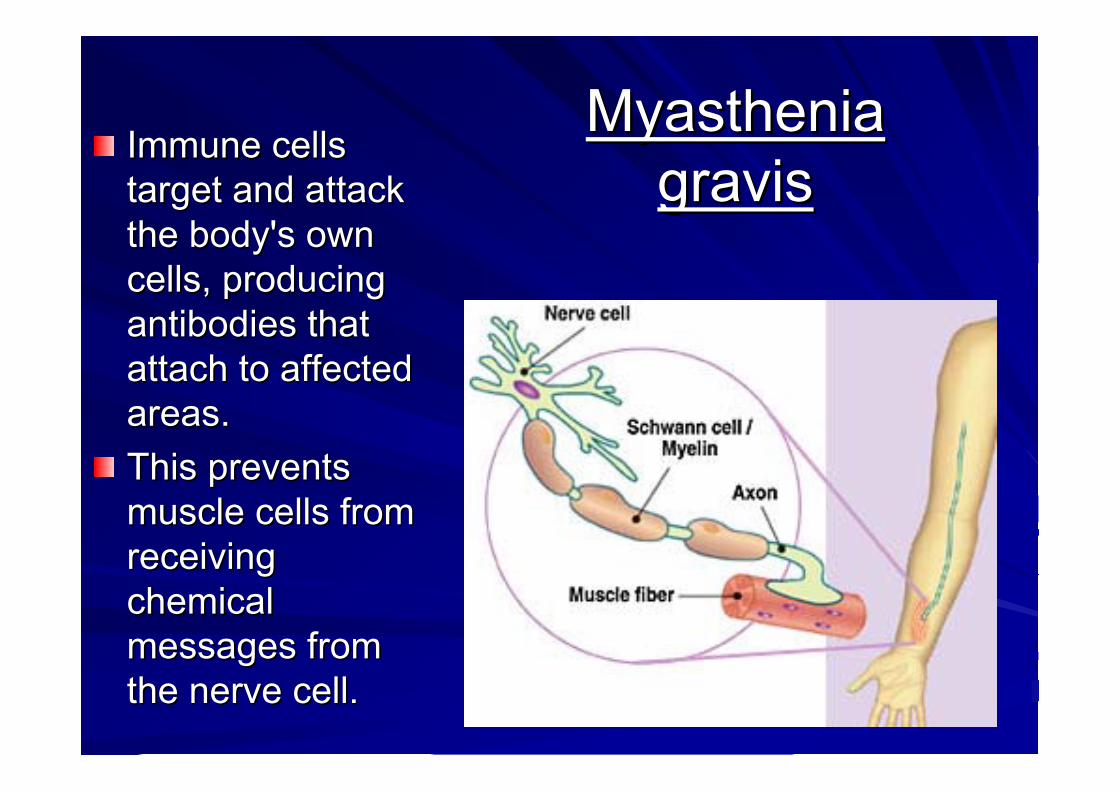
Myasthenia Myasthenia gravisgravis
Immune cells Immune cells target and attack target and attack the body's own the body's own cells, producing cells, producing antibodiesantibodies that that attach to affected attach to affected areas. areas. This prevents This prevents muscle cells from muscle cells from receiving receiving chemical chemical messages from messages from the nerve cell.the nerve cell.

Myasthenia GravisMyasthenia Gravis
Eyelids can droopEyelids can droop
Eye muscles can no longer coordinate right & left Eye muscles can no longer coordinate right & left eye synchronous movement, so double vision eye synchronous movement, so double vision often occursoften occurs
Chewing & swallowing becomes difficultChewing & swallowing becomes difficult
Arms & legs can be affectedArms & legs can be affected

Patients with Patients with myasthenia myasthenia gravis have a gravis have a higher risk of higher risk of having other having other autoimmune autoimmune disorders. disorders. The cause of The cause of autoimmune autoimmune disordersdisorders is is unknown. unknown.
Myasthenia Myasthenia gravisgravis

Myasthenia GravisMyasthenia GravisClinical ManifestationsClinical Manifestations
Primary features is increasing weakness with sustained Primary features is increasing weakness with sustained muscle contractionmuscle contraction
Extreme muscle weakness and generalized fatigueExtreme muscle weakness and generalized fatigueIncrease fatigueIncrease fatigue--acethycholineacethycholine taken or used taken or used
up by movement, pt gets weaker & weaker. While at rest, up by movement, pt gets weaker & weaker. While at rest, not using as much not using as much acethycholineacethycholine..–– Worse following effortWorse following effort–– Relieved with restRelieved with rest
Symptoms vary according to muscles involvedSymptoms vary according to muscles involved

Myasthenia GravisMyasthenia GravisClinical ManifestationsClinical Manifestations
Symmetric muscles involved, especially Symmetric muscles involved, especially those innervated by cranial nervesthose innervated by cranial nerves–– DiplopiaDiplopia --double visiondouble vision–– PtosisPtosis--droopy eyeliddroopy eyelid–– Sleepy mask like expressionSleepy mask like expression–– DysphoniaDysphonia--diff speaking, hoarsenessdiff speaking, hoarseness–– DysphagiaDysphagia--inability or difficulty in swallowinginability or difficulty in swallowing
tendency for mouth to hang opentendency for mouth to hang open–– Progressive weakness of diaphragm/intercostalsProgressive weakness of diaphragm/intercostals–– Variable course with Variable course with excerbationexcerbation’’ss/remissions/remissions

Myasthenia GravisMyasthenia GravisMotor & Sensory Clinical ManifestationsMotor & Sensory Clinical Manifestations
Motor ManifestationsMotor ManifestationsProgressive muscle weakness (proximal) that usually Progressive muscle weakness (proximal) that usually improves with restimproves with restPoor posturePoor postureOcular palsiesOcular palsiesPtosisPtosis/Weak or incomplete eye closure/Weak or incomplete eye closureDiplopiaDiplopiaRespiratory compromise Respiratory compromise secondary to ineffective secondary to ineffective coughing, swallowing, muscle weakness, etc.coughing, swallowing, muscle weakness, etc.Loss of bowel & bladder controlLoss of bowel & bladder controlSensory ManifestationSensory ManifestationMuscle achinessMuscle achinessParesthesiasParesthesiasDecreased smell and tasteDecreased smell and taste

DxDx of Myasthenia gravisof Myasthenia gravisHistory & PhysicalHistory & PhysicalTensilonTensilon TestingTesting -- Confirmed by Confirmed by injection of injection of anticholinesteraseanticholinesterase drugsdrugs, usually , usually TensilonTensilon((edrophoniumedrophonium) & patients response to cholinergic drugs ) & patients response to cholinergic drugs (see next slides)(see next slides)Lab StudiesLab Studies–– Thyroid function testedThyroid function tested–– Serum protein electrophoresis evaluates pt for Serum protein electrophoresis evaluates pt for imunologicimunologic
disorderdisorder–– ThyrotoxicosisThyrotoxicosis (excessive thyroid hormone) is present in approx (excessive thyroid hormone) is present in approx
5% of MG patients5% of MG patientsOther Other dxdx associated with MGassociated with MG --Rheumatoid arthritis, Rheumatoid arthritis, systemic lupus systemic lupus erythematosuserythematosus, & , & polymyositispolymyositisEMG EMG -- ElectromyographyElectromyographyElectrical testing of normal neuromuscular junction should Electrical testing of normal neuromuscular junction should produce no change in the amplitude of muscle contraction. In produce no change in the amplitude of muscle contraction. In MG, the amplitude of the muscleMG, the amplitude of the muscle’’s response diminishes with s response diminishes with progressive stimulation = MGprogressive stimulation = MG

Myasthenia Gravis Myasthenia Gravis -- CrisisCrisis
Both types of CrisisBoth types of Crisis --Sudden increase in Sudden increase in weakness & inability to clear secretions, weakness & inability to clear secretions, swallow, or breath adequately. Patient will swallow, or breath adequately. Patient will choke on their own secretionschoke on their own secretions
2 types of Crisis2 types of Crisis::MyasthenicMyasthenic Gravis CrisisGravis Crisis
Cholinergic CrisisCholinergic Crisis
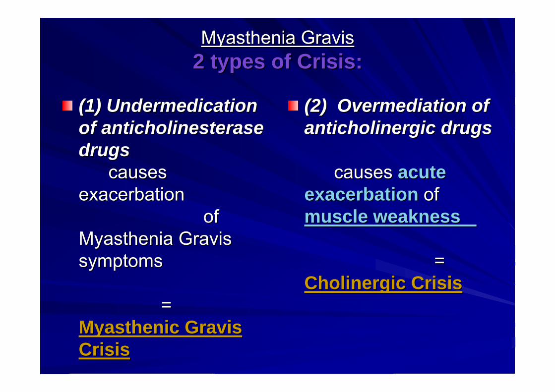
Myasthenia GravisMyasthenia Gravis2 types of Crisis:2 types of Crisis:
(1) (1) UndermedicationUndermedicationof of anticholinesteraseanticholinesterasedrugsdrugs
causes causes exacerbation exacerbation
of of Myasthenia Gravis Myasthenia Gravis symptomssymptoms
= = MyasthenicMyasthenic Gravis Gravis CrisisCrisis
(2) (2) OvermediationOvermediation of of anticholinergicanticholinergic drugsdrugs
causes causes acute acute exacerbationexacerbation of of muscle weaknessmuscle weakness
= = Cholinergic CrisisCholinergic Crisis

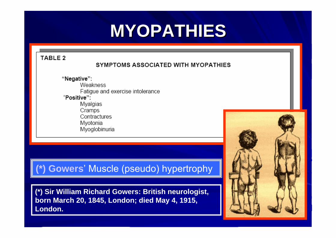
MYOPATHIESMYOPATHIES
MYOPATHIESMYOPATHIES
(*) Gowers’ Muscle (pseudo) hypertrophy
(*) Sir William Richard Gowers: British neurologist, born March 20, 1845, London; died May 4, 1915, London.

(*) Gowers' sign
(*) Standing up with the aid of hands pushing on kneesSir William Richard Gowers: British neurologist, bornMarch 20, 1845, London; died May 4, 1915, London.
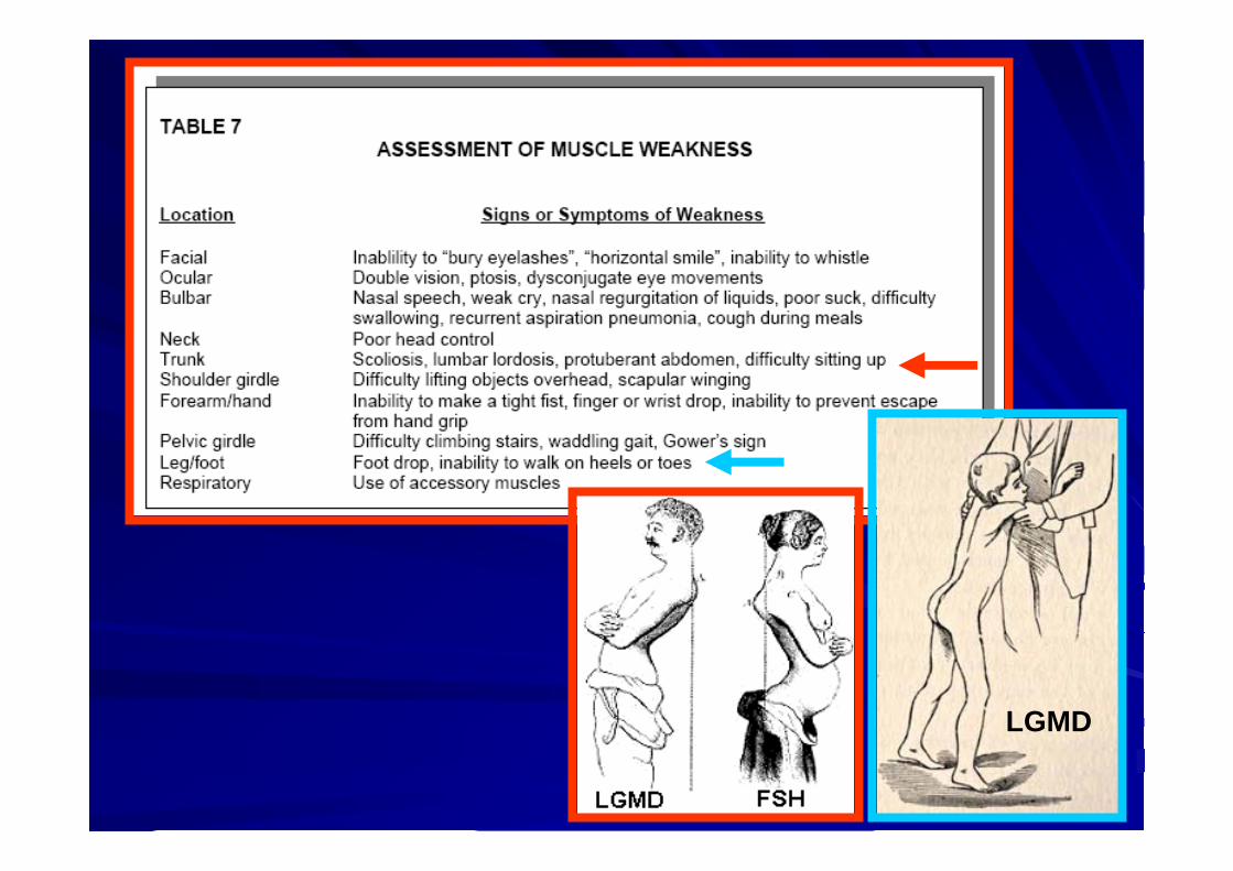
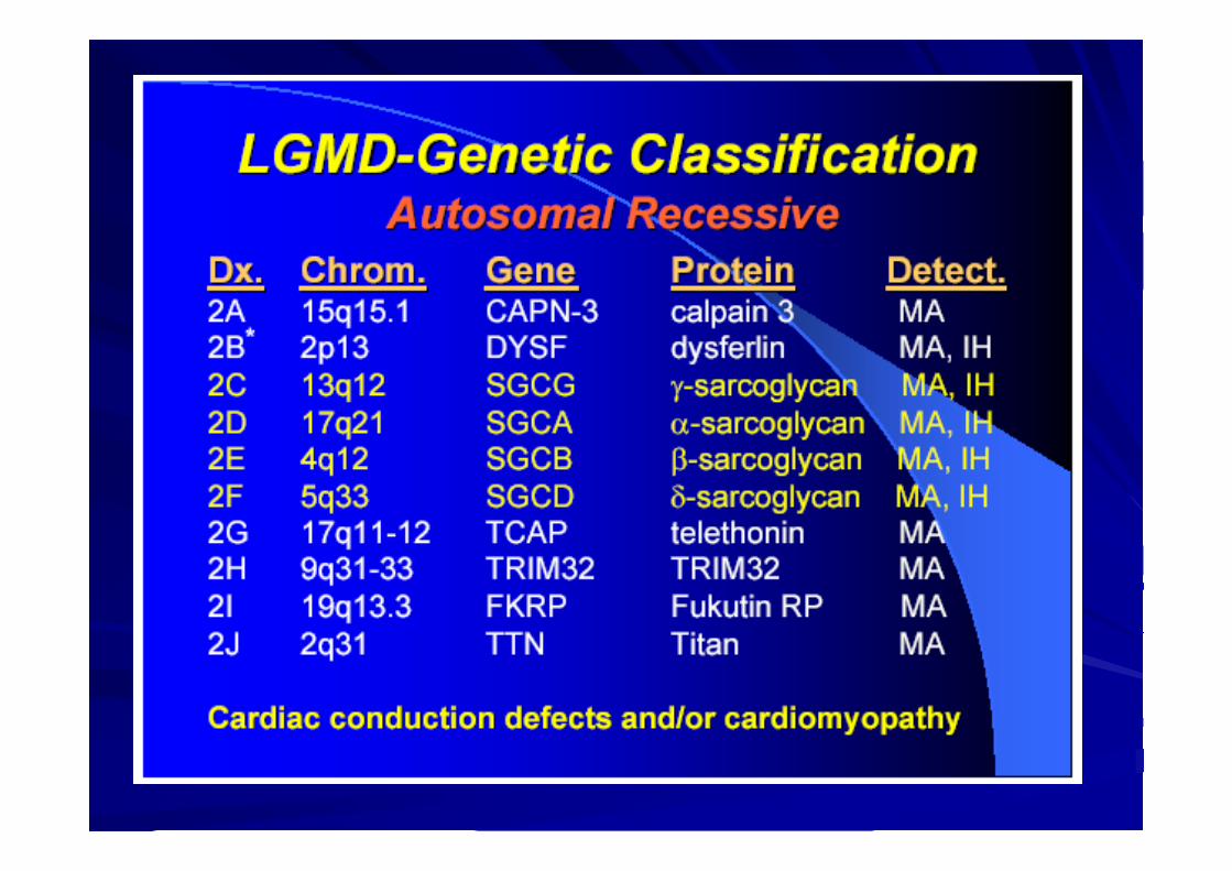
LGMD

DYSTROPHINOPATHIESDYSTROPHINOPATHIES
Guillaume-BenjaminDuchenne de Boulogne
Autographed copy of: De la Paralysie Musculaire Pseudo-hypertrophique...1868
Duchenne muscular dystrophy

Duchenne Muscular Distrophy is unique in being the only musculardystrophy for which a therapy (prednisone and deflazacort) has beenproven effective in randomized, controlled trials.
(Live birth)

DuchenneDuchennemuscularmusculardystrophydystrophy
ChromosomeChromosome Xp21; RecessiveXp21; RecessiveOnsetOnset 3 3 toto 5 5 yrsyrsClinicalClinical–– WeaknessWeakness DistributionDistribution
ProximalProximal > > DistalDistalSymmetricSymmetricLegsLegs & & ArmsArmsMostMost involvedinvolved musclesmuscles: : AdductorAdductor magnusmagnus in in legslegsRelativelyRelatively sparedspared musclesmuscles: : GracilisGracilis & & SartoriusSartorius
-- CourseCourseReducedReduced motor motor functionfunction byby 2 2 toto 3 3 yearsyearsSteady Steady declinedecline in in strengthstrength: After 6 : After 6 toto 11 11 yearsyearsGowersGowers signsignFailureFailure toto walkwalk: 9 : 9 -- 13 13 yearsyears; ; LaterLater withwith steroidsteroid treatmenttreatment
Standing from supine position

DuchenneDuchenne muscularmusculardystrophydystrophy
• Muscle (pseudo)hypertrophy
Especially calfMay be generalizedIncreases with ageMost commonly due tomuscle fibrosisSome relatively sparedmuscles may have truehypertrophy
macroglossia
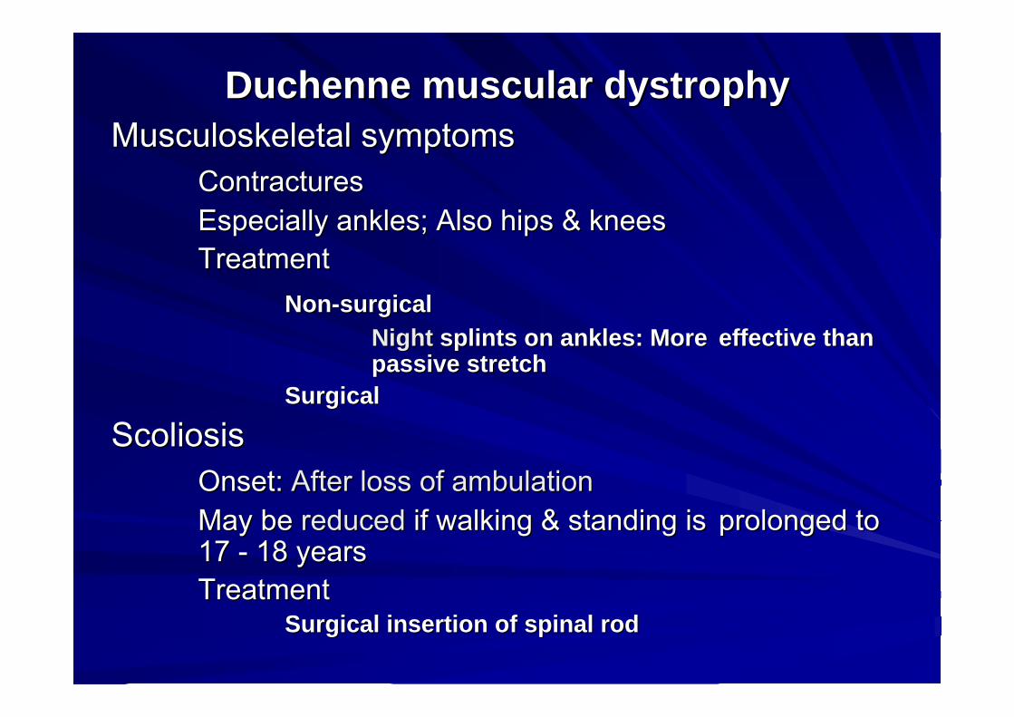
DuchenneDuchenne muscularmuscular dystrophydystrophyMusculoskeletalMusculoskeletal symptomssymptoms
ContracturesContracturesEspeciallyEspecially anklesankles; ; AlsoAlso hipships & & kneeskneesTreatment Treatment
NonNon--surgicalsurgicalNight Night splintssplints on on anklesankles: More : More effectiveeffective thanthanpassive stretch passive stretch
SurgicalSurgical
ScoliosisScoliosisOnsetOnset: : After loss of After loss of ambulationambulationMay May bebe reducedreduced ifif walkingwalking & standing & standing isis prolongedprolonged toto17 17 -- 18 18 yearsyearsTreatmentTreatment
SurgicalSurgical insertioninsertion of of spinalspinal rodrod

DuchenneDuchenne muscularmuscular dystrophydystrophyOtherOther clinicalclinical featuresfeatures
CardiomyopathyCardiomyopathy: : DilatedDilated; ; EspeciallyEspecially > 15 > 15 yearsyearsMentalMental retardationretardation: : MeanMean IQ ~ 88 IQ ~ 88 Night Night blindnessblindnessAlteredAltered responseresponse toto flashesflashes of light in dark of light in dark adaptedadaptedstate state ERG: ERG: bb--wavewave, , ReducedReduced amplitudeamplitudeDp260: Dp260: IsoformIsoform of of dystrophindystrophin in retina in retina
• Death Most common between 15 - 25 yearsDue to respiratory or cardiac failureLife prolonged by ~ 6 years to 25 years withrespiratory supportLife shortened by 2 years with cardiomyopathy
Reduced verbal IQ Selective defects: - Shorter digit span memory- Developmental delay
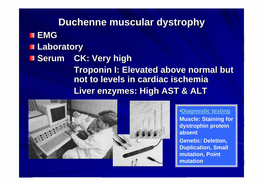
DuchenneDuchenne muscularmuscular dystrophydystrophyEMGEMGLaboratoryLaboratorySerumSerum CK: CK: VeryVery high high
TroponinTroponin I: I: ElevatedElevated aboveabove normalnormal butbutnotnot toto levelslevels in in cardiaccardiac ischemiaischemiaLiverLiver enzymesenzymes: High AST & ALT : High AST & ALT
•Diagnostic testingMuscle: Staining fordystrophin proteinabsentGenetic: Deletion, Duplication, Smallmutation, Pointmutation

Duchenne musculardystrophy
• Muscle biopsy: General featuresVariable fiber size: Small fibers roundedHypercontracted (opaque) muscle fbersNecrosis “Myopathic grouping”Muscle fiber degeneration & regeneration:Especially early
Muscle fiber internal archetecture: Normal or immature

Duchennemusculardystrophy
• Dystrophin: Absent staining• Other membrane proteins
• Sarcoglycans: Reduced• Aquaporin 4: Reduced
• Endomysial fibrosis: More with late pathology
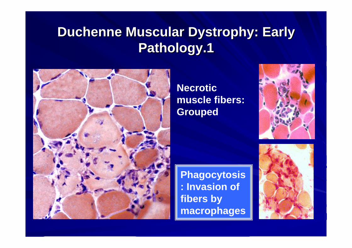
DuchenneDuchenne MuscularMuscular DystrophyDystrophy: : EarlyEarlyPathologyPathology.1.1
Necroticmuscle fibers: Grouped
Phagocytosis: Invasion of fibers bymacrophages

DuchenneDuchenne MuscularMuscular DystrophyDystrophy: : EarlyEarlyPathologyPathology. 2. 2
Immature muscle fibers: Numerous
Myopathic grouping of regenerating muscle fibers
Many smallregenerating fiber
Intermediate-sized regeneratingmuscle fibers: Myopathic grouping
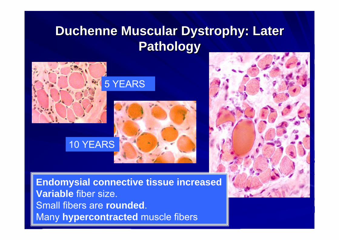
DuchenneDuchenne MuscularMuscular DystrophyDystrophy: : LaterLaterPathologyPathology
5 YEARS
10 YEARS
Endomysial connective tissue increasedVariable fiber size.Small fibers are rounded.Many hypercontracted muscle fibers

DuchenneDuchenne MuscularMuscularDystrophyDystrophy:: DystrophinDystrophin stainingstaining
Normal dystrophinstainingaround the rim of muscle fibers
Absent dystrophin: Duchennemuscular dystrophyLeft: No staining around the rim of muscle fibers.Right: No staining of most musclefibers. One "revertant" fiber withdystrophin staining.
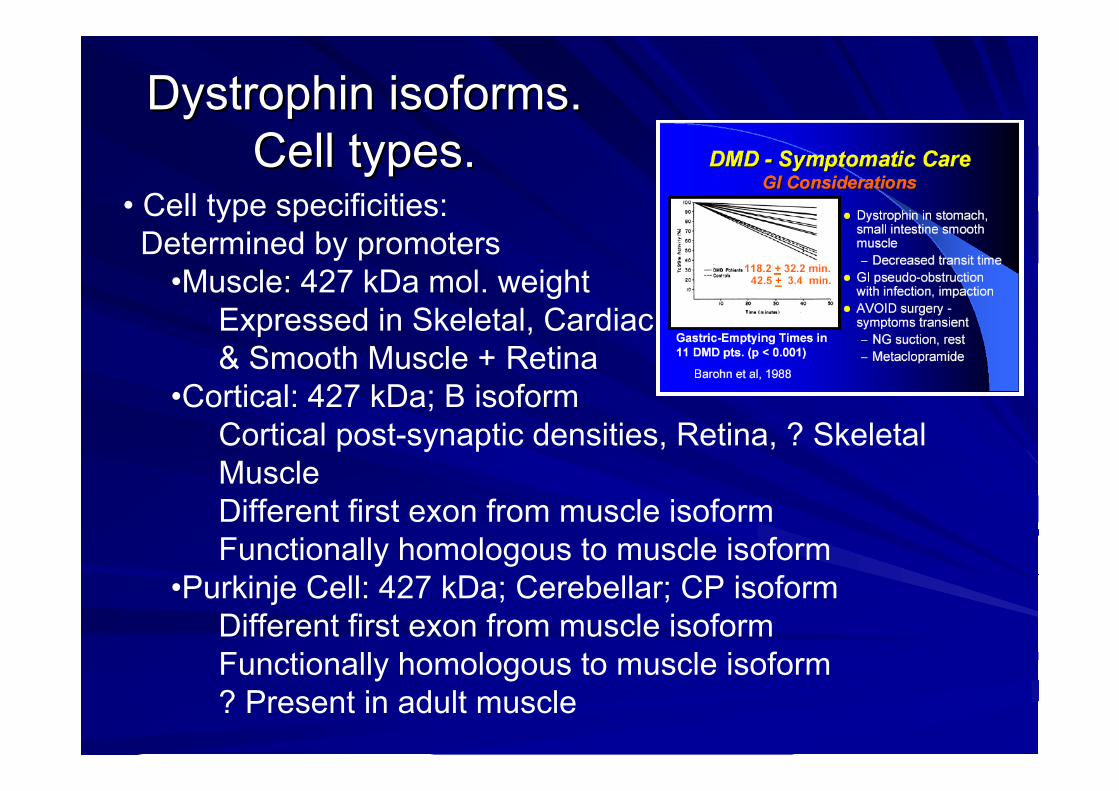
DystrophinDystrophin isoformsisoforms. . CellCell typestypes..
• Cell type specificities: Determined by promoters
•Muscle: 427 kDa mol. weightExpressed in Skeletal, Cardiac& Smooth Muscle + Retina
•Cortical: 427 kDa; B isoformCortical post-synaptic densities, Retina, ? SkeletalMuscleDifferent first exon from muscle isoformFunctionally homologous to muscle isoform
•Purkinje Cell: 427 kDa; Cerebellar; CP isoformDifferent first exon from muscle isoformFunctionally homologous to muscle isoform? Present in adult muscle

•Retinal: 260 kDaRetinal exon 1 spliced to exon 30 Mouse retina: outer plexiform layer
•Brain (Fetal) & Kidney: 140 kDaPromoter & first exon: In large intron between exon 44 and 45
•Schwann cell (S-dystrophin): 116 kDaOnset exon 56 Submembrane of external (abaxonal) layer; Nodes of RanvierMouse model: Reduced dystrophin in peripheral nervecauses demyelinating neuropathy
•Glial: 71 kDaOnset exon 63 Brain (Glia), Viscera (Lung, Liver, Kidney), Cardiac Muscle
DystrophinDystrophin localizationlocalization. . CellCell typestypes..

•Toewalking
•Enlargedcalves
•Clinical features• Onset > 7 yrs• Weakness
Proximal > Distal; Symmetric;Legs & ArmsMay be especiallyprominent in quadricepsor hamstringsSlowly progressive Calf pain on exercise
• Muscle hypertrophy: Especially calves
• Failure to walk 16 – 80years
BeckerBecker muscularmuscular dystrophydystrophy

• Genotype: Dystrophin mutations•Deletion 70% of patients: Usually In-frame
16% with frameshift mutationPoint mutations
> 70 identified; new mutation rare •Worse course: Additional mutation in Myogenic factor 6
• Systemic• Joint contractures: Ankles & Other• Cardiomyopathy: May occur before severe weakness• Mental retardation
- Associated with deletion of Dp140 transcription unit• Serum CK
• Very high: 5,000 to 20,000 • Lower levels with increasing age & disability
BeckerBecker muscularmuscular dystrophydystrophy
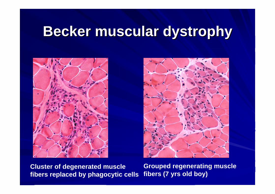
BeckerBecker muscularmuscular dystrophydystrophy
Cluster of degenerated musclefibers replaced by phagocytic cells
Grouped regenerating musclefibers (7 yrs old boy)

BeckerBecker muscularmuscular dystrophydystrophy
• chronic dystrophy:•Increased endomysial connective tissue•Variable fiber size: Small muscle fibers are rounded•Internal nuclei •The largest muscle fibers are hypertrophied•Occasional fibers: Degeneration; Regeneration; Hypercontraction; Split
27 yrs old male

BeckerBecker muscularmuscular dystrophydystrophy
Normal dystrophinstainingaround the rim of musclefibers
Reduced dystrophinstainingSeverity & onset agecorrelate with muscledystrophin levels
Dystrophin staining

DystrophinopathiesDystrophinopathies: : CardiomyopathyCardiomyopathy• Cardiomyopathy with Becker or Duchenne MD syndromes: Common
Common mutations: Deletions in Exons 48-53 region(Spectrin-like region) Clinical features
Tachycardia: > 100; Common even < 10 years ageDilated cardiomyopathySymptomatic: 57% by age 18 Progression: VariableMay be associated with only: Myalgias after exercise & High CK
EKG Changes due to atrophyAge 10: EKG changes in 60%; No clinical
manifestations. Age 18: EKG or ECHO changes in most
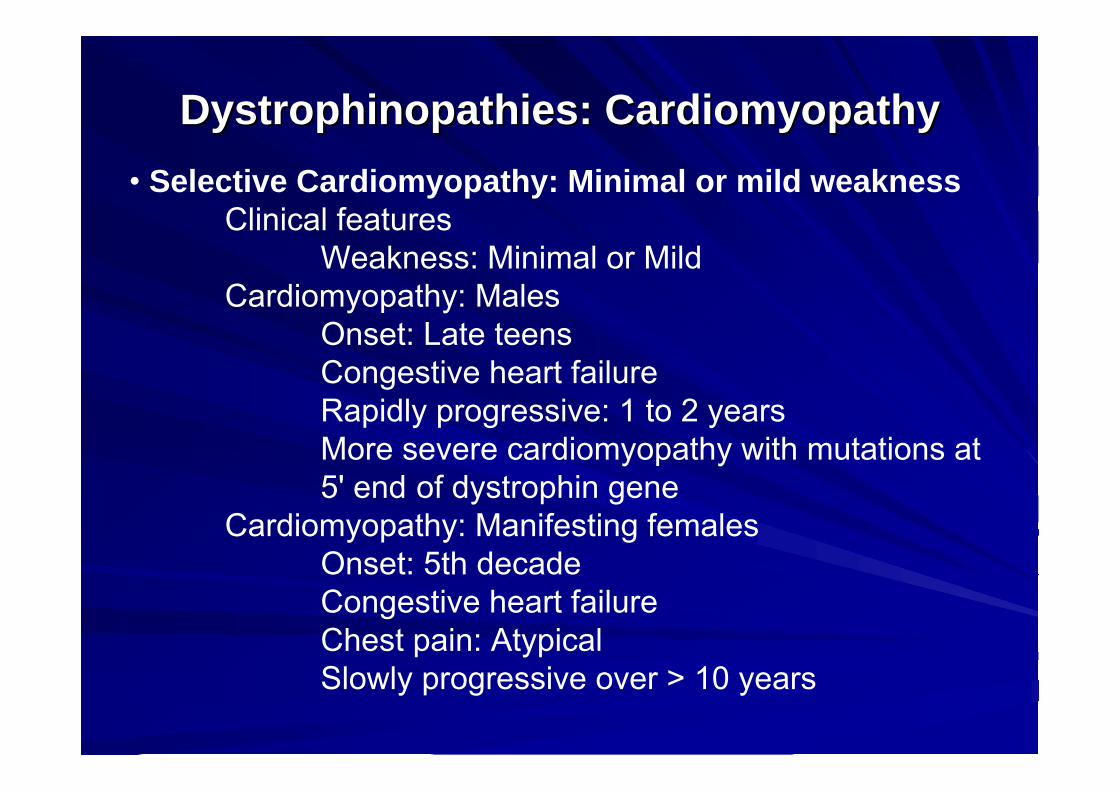
DystrophinopathiesDystrophinopathies: : CardiomyopathyCardiomyopathy• Selective Cardiomyopathy: Minimal or mild weakness
Clinical featuresWeakness: Minimal or Mild
Cardiomyopathy: MalesOnset: Late teensCongestive heart failureRapidly progressive: 1 to 2 yearsMore severe cardiomyopathy with mutations at 5' end of dystrophin gene
Cardiomyopathy: Manifesting femalesOnset: 5th decade Congestive heart failureChest pain: AtypicalSlowly progressive over > 10 years
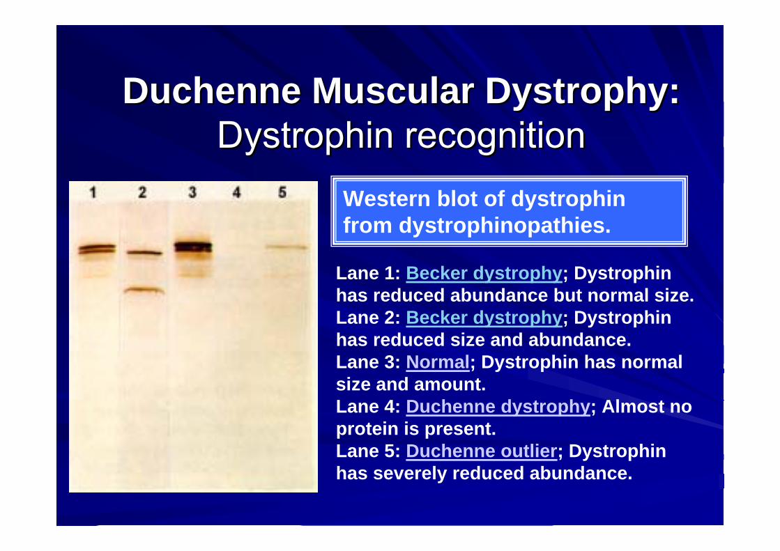
DuchenneDuchenne MuscularMuscular DystrophyDystrophy::DystrophinDystrophin recognitionrecognition
Lane 1: Becker dystrophy; Dystrophinhas reduced abundance but normal size.Lane 2: Becker dystrophy; Dystrophinhas reduced size and abundance.Lane 3: Normal; Dystrophin has normalsize and amount.Lane 4: Duchenne dystrophy; Almost no protein is present.Lane 5: Duchenne outlier; Dystrophinhas severely reduced abundance.
Western blot of dystrophinfrom dystrophinopathies.
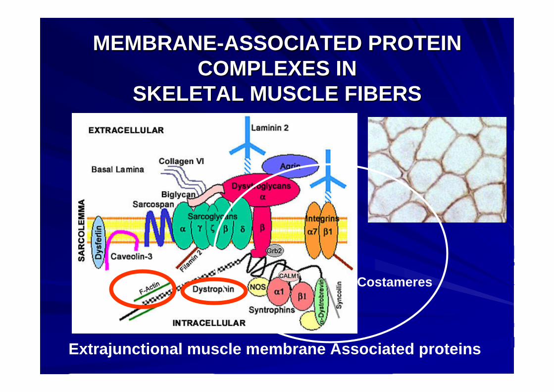
MEMBRANEMEMBRANE--ASSOCIATED PROTEIN ASSOCIATED PROTEIN COMPLEXES INCOMPLEXES IN
SKELETAL MUSCLE FIBERS SKELETAL MUSCLE FIBERS
Extrajunctional muscle membrane Associated proteins
Costameres
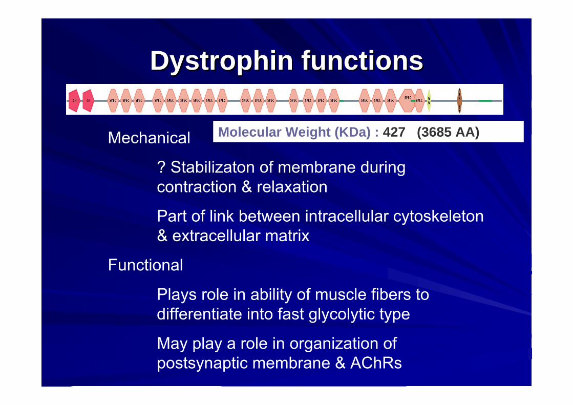
DystrophinDystrophin functionsfunctions
Mechanical
? Stabilizaton of membrane duringcontraction & relaxation
Part of link between intracellular cytoskeleton& extracellular matrix
Functional
Plays role in ability of muscle fibers todifferentiate into fast glycolytic type
May play a role in organization of postsynaptic membrane & AChRs
Molecular Weight (KDa) : 427 (3685 AA)
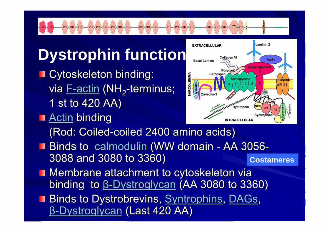
CytoskeletonCytoskeleton bindingbinding: : via via FF--actinactin (NH(NH22--terminus;terminus;1 1 stst toto 420 AA)420 AA)ActinActin bindingbinding(Rod: (Rod: CoiledCoiled--coiledcoiled 2400 amino 2400 amino acidsacids))BindsBinds toto calmodulincalmodulin (WW domain (WW domain -- AA 3056AA 3056--3088 and 3080 3088 and 3080 toto 3360)3360)Membrane attachment Membrane attachment toto cytoskeletoncytoskeleton via via bindingbinding toto ββ--DystroglycanDystroglycan (AA 3080 (AA 3080 toto 3360)3360)BindsBinds toto DystrobrevinsDystrobrevins, , SyntrophinsSyntrophins, , DAGsDAGs, , ββ--DystroglycanDystroglycan (Last 420 AA) (Last 420 AA)
Dystrophin functions
Costameres

• Ca++ ions release fromsarcoplasmic reticulum isregulated by 2 large membrane protein complexes:• Dihydropyridine receptor
(DHPR)• Ryanodyne receptor (RyR)
Action potential• travels along muscle
surface membrane • enters transverse
tubule system (t-tubules)
Costameres
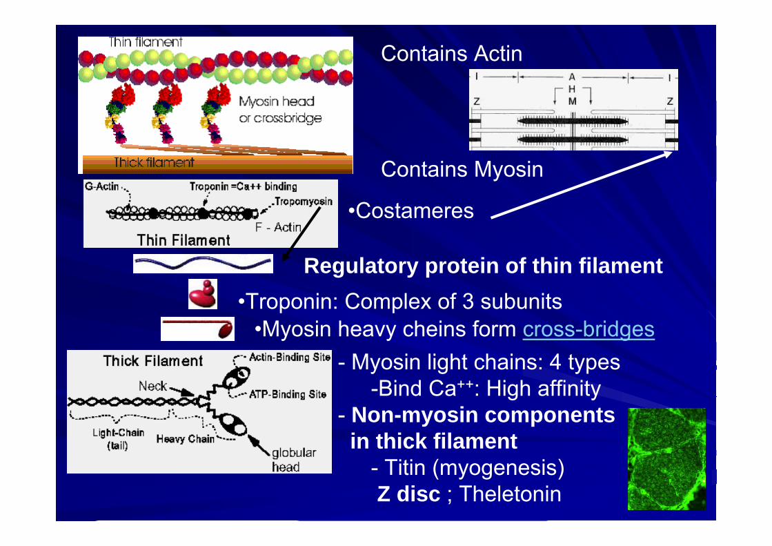
Contains Myosin
Contains Actin
- Myosin light chains: 4 types-Bind Ca++: High affinity
- Non-myosin componentsin thick filament
- Titin (myogenesis)Z disc ; Theletonin
•Troponin: Complex of 3 subunitsRegulatory protein of thin filament
•Myosin heavy cheins form cross-bridges
•Costameres

Myosin
Actin
Contractile apparatus activated• Ca++ binds to troponin complex• Tropomyosin binding to contractile apparatus changes• Actin allowed to bind to myosin heads• Muscle contraction occurs via myofilament sliding

DystrophinDystrophin gene gene mutationsmutations: : SizeSize & & TypesTypesPoint Point mutationsmutations alongalong entireentire genegeneOftenOften cause premature cause premature truncationtruncation of of translationtranslationMay May occuroccur withwith DuchenneDuchenne or Becker or Becker phenotypephenotypeFrequencyFrequency: : DetectedDetected in ~73% of in ~73% of patientspatients withoutwithout deletionsdeletions or or duplicationsduplications
DeletionsDeletions or or DuplicationsDuplicationsMajority of Majority of deletionsdeletions at the 3' end at the 3' end regionregion; 5' end ; 5' end deletionsdeletions in 18% in 18% More More thanthan one one exonexon usuallyusually deleteddeletedClinicalClinical correlationscorrelations::
ExonExon 1 & promoter 1 & promoter regionregion: : MildMild weaknessweakness ±± severeseverecardiomyopathycardiomyopathyDeletionsDeletions of more of more thanthan 36 36 exonsexons produce severe produce severe phenotypephenotype
DiseaseDisease frequencyfrequencyDuchenneDuchenne: 65% of : 65% of patientspatients havehave gene gene deletionsdeletionsBecker: 55% Becker: 55% toto 85% of 85% of patientspatients havehave gene gene deletionsdeletions or or duplicationsduplications
oo VeryVery largelarge chromosomalchromosomal deletionsdeletionsMultisystemMultisystem disordersdisordersDuchenneDuchenne muscularmuscular dystrophydystrophy phenotypephenotype
DystrophinDystrophinabnormalitiesabnormalities

EffectsEffects of of dystrophindystrophin mutationsmutations: : OtherOtherDystrophinDystrophin absenceabsence
DMD DMD phenotypephenotype: Severe : Severe ReductionReduction in in sarcoglycanssarcoglycans & & otherother proteinsproteins in in dystrophindystrophin--glycoproteinglycoprotein complexcomplexDysferlinDysferlin increasedincreased in in cytoplasmcytoplasm
CC--terminal terminal mutationsmutationsSevere Severe dystrophiesdystrophies
FunctionalFunctional consequencesconsequences of loss of of loss of dystrophindystrophin on on musclemusclefibersfibers
IncreasedIncreased movementmovement of membrane of membrane impermeantimpermeantmoleculesmolecules intointo and out of and out of musclemuscle cellscellsForce production: Force production: DecreasedDecreased; ; HypersensitiveHypersensitive totolengtheninglengthening, or , or eccentriceccentric contractioncontractionForce Force decrementdecrement withwith eccentriceccentric contractioncontraction correlatescorrelateswithwith acutelyacutely increasedincreased sarcolemmalsarcolemmal permeabilitypermeabilityDisorganizedDisorganized subsarcolemmalsubsarcolemmal costamerescostameres
DystrophinDystrophinabnormalitiesabnormalities

LimbLimb--GirdleGirdle MuscularMuscular DystrophDystrophiesiesThe term “limb-girdle muscular dystrophy”(LGMD) refers not to a single disease, butrather to group of disorders, all of whichusually but, paradoxically, not always, involve mainly proximal muscles.
In fact, most LGMD patients have weaknessbeyond a simple limb-girdle distribution(LGMD 2B).
15 forms (and their genetic defect; 5 dominant; 10 recessive) have been identified)


MYOPATHIES WITH PTOSIS MYOPATHIES WITH PTOSIS +/+/-- OPHTALMOPLEGIAOPHTALMOPLEGIA
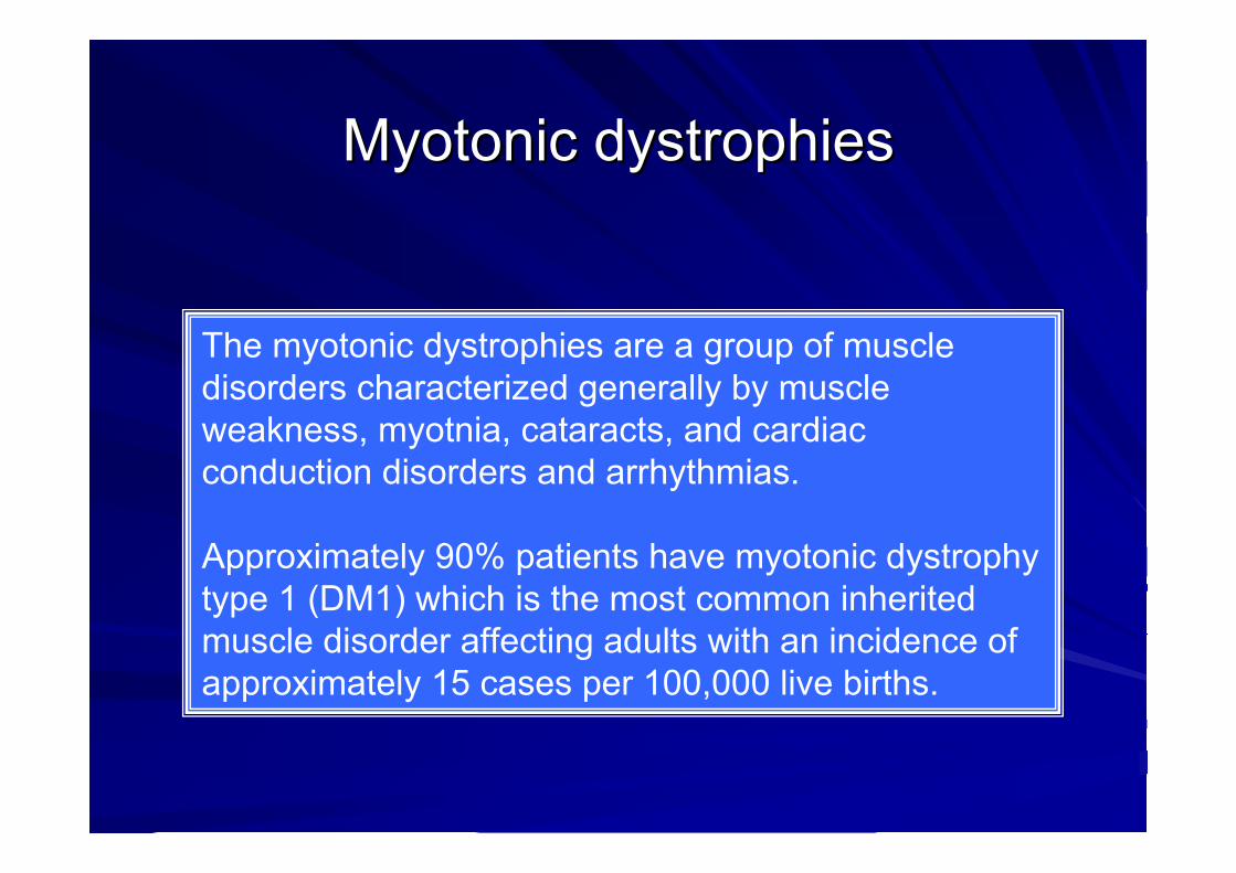
MyotonicMyotonic dystrophdystrophiesies
The myotonic dystrophies are a group of muscledisorders characterized generally by muscleweakness, myotnia, cataracts, and cardiacconduction disorders and arrhythmias.
Approximately 90% patients have myotonic dystrophytype 1 (DM1) which is the most common inheritedmuscle disorder affecting adults with an incidence of approximately 15 cases per 100,000 live births.

2
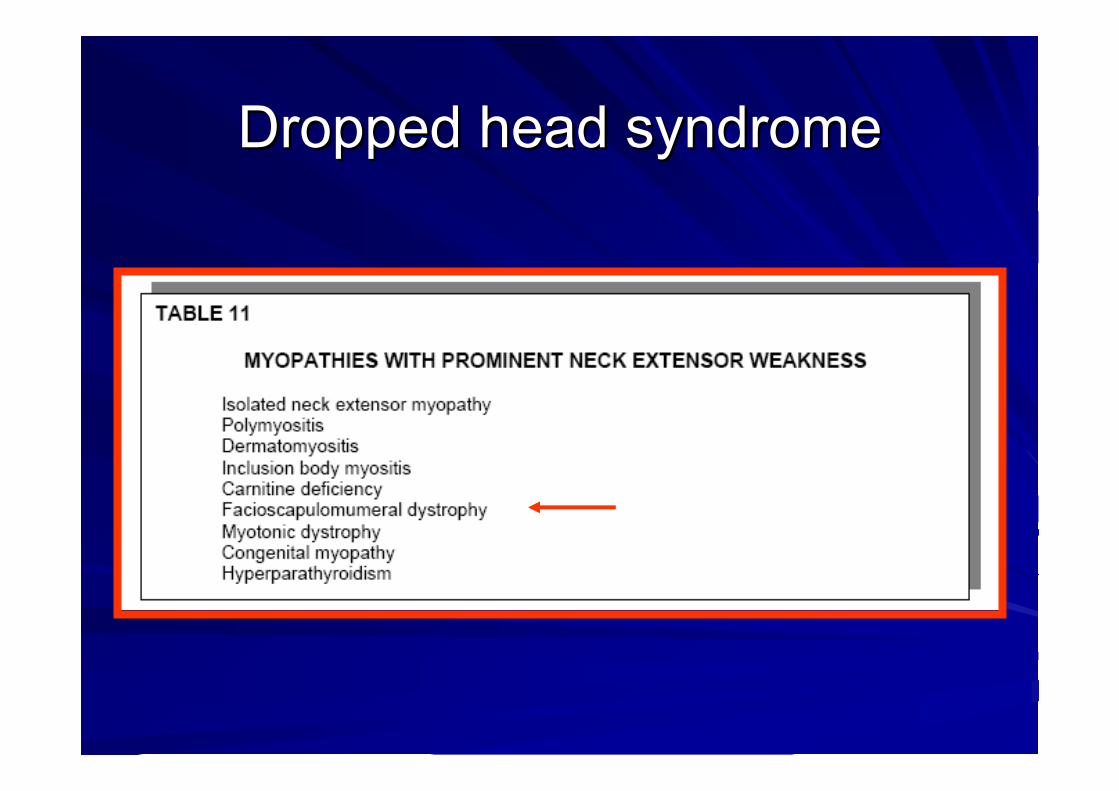
DroppedDropped head head syndromesyndrome

FacioscapulohumeralFacioscapulohumeral DystrophyDystrophy
Although FSHD was described over a century ago, and is the third most common dystrophy prevalence1:20,000), it has received relatively little attention.
It appears that FSHD results from inappropriate over-expression of certain genes, rather than the absence or under-expression of genes, as in most dystrophies.
Mechanisms of toxic effect on muscle cell are unknown


DISTAL WEAKNESS IN DISTAL WEAKNESS IN MYOPATHIESMYOPATHIES

DISTAL WEAKNESS IN DISTAL WEAKNESS IN MYOPATHIESMYOPATHIES
Chromosome 2p13; Dominant•Genetics10
•Not allelic with Miyoshi myopathy & LGMD 2B•Finnish & Swedish patients have shared haplotype
•Epidemiology: Especially mid-Sweden & Finland •Onset
•Age: Usually > 40 years; Median 5th decade; Range 20 to 77 •Location: Arms; Wrist & Finger extensors
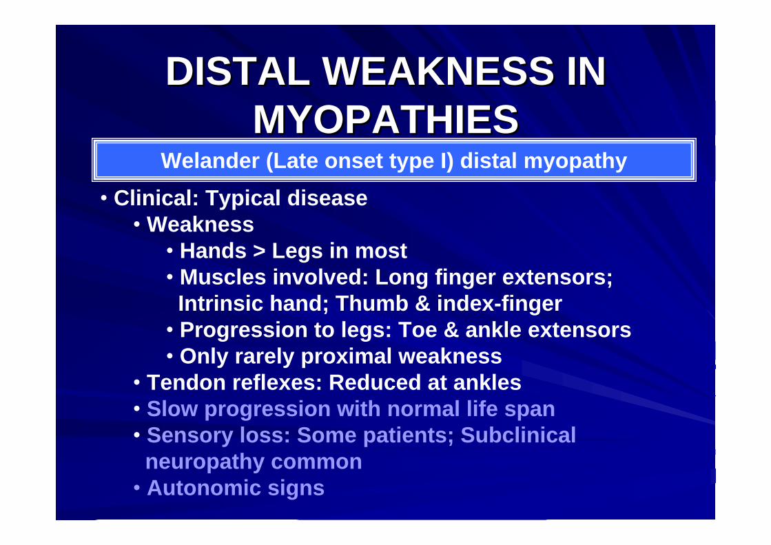
Welander (Late onset type I) distal myopathy
DISTAL WEAKNESS IN DISTAL WEAKNESS IN MYOPATHIESMYOPATHIES
• Clinical: Typical disease• Weakness
• Hands > Legs in most• Muscles involved: Long finger extensors;Intrinsic hand; Thumb & index-finger
• Progression to legs: Toe & ankle extensors• Only rarely proximal weakness
• Tendon reflexes: Reduced at ankles• Slow progression with normal life span• Sensory loss: Some patients; Subclinicalneuropathy common
• Autonomic signs
Welander (Late onset type I) distal myopathy

DISTAL WEAKNESS IN DISTAL WEAKNESS IN MYOPATHIESMYOPATHIES
• Laboratory•CK: Normal or mildly elevated•EMG: Myopathic; Some irritability•Muscle Pathology•Chronic myopathic: Varied fiber size; Splitting•Rimmed vacuoles: Variably present•Tubulo-Filamentous inclusions: Sarcoplasm & muscle fiber nuclei•Eosinophilic cytoplasmic bodies•Neurogenic changes
Welander (Late onset type I) distal myopathy


