
UNIVERSITA’ DEGLI STUDI DI PADOVA
DIPARTIMENTO DI BIOLOGIA
SCUOLA DI DOTTORATO DI RICERCA IN : BIOSCIENZE E BIOTECNOLOGIE
INDIRIZZO: NEUROBIOLOGIA
CICLO: XXVI
ROLE OF AUTOPHAGY IN AGE-RELATED MUSCLE LOSS
Direttore della Scuola: Ch.mo Prof. Giuseppe Zanotti
Coordinatore d’indirizzo: Ch.ma Prof.ssa Daniela Pietrobon
Supervisore: Prof. Marco Sandri
Dottoranda: Francesca Lo Verso

2
3
INDEX
Riassunto…………………………………………………………………………………………………………. 7
Summary………………………………………………………………………………………………………….13
1. INTRODUCTION
1.1 Skeletal muscle………………………………………………………………………………. 17
1.1.1 Structure and function…………………………………………………………..17
1.1.2 The nerve-muscle connection……………………………………………….. 12
1.2 Muscle hypertrophy and atrophy ……………………………………………………. 28
1.3 Ageing in muscle tissue: sarcopenia………………………………………………… 30
1.4 The autophagy-lysosomal system……………………………………………………. 35
1.4.1 The autophagy genes……………………………………………………………. 37
1.4.2 Autophagy machinery………………………………………………………….. 38
1.4.3 Mitophagy…………………………………………………………………………… 42
1.4.4 Molecular signalling in autophagy ……………………………………….. 44
1.4.5 Autophagy in disease…………………………………………………………… 49
1.5 Autophagy and muscle……………………………………………………………………. 49
1.5.1 Regulation of autophagy in skeletal muscle…………………………… 49
1.5.2 The in vivo model of muscle-specific block of autophagy…………51
1.6 Autophagy and ageing ……………………………………………………………………..54
1.7 Autophagy and exercise………………………………………………………………….. 55
1.8 Aim of the work………………………………………………………………………………. 57
2. MATERIALS AND METHODS
2.1 Generation of muscle-specific Atg7 knockout mice…………………………… 59
2.1.1 Genotyping of muscle specific Atg7 knockout mice......................... 59
2.2 In vivo skeletal muscle electroporation……………………………………………..60
2.3 Measurements of muscle force in vivo……………………………………………… 61
2.4 Histology analyses and fibre size measurement……………………………….. 62
2.4.1 Haematoxylin and Eosin staining (H&E) ………………………………. 62
2.4.2 Succinate dehydrogenase (SDH) …………………………………………. 63
4
2.4.3 Fibre cross-sectional area (CSA) ………………………………………….. 63
2.5 Immunoistochemistry analyses……………………………………………………….. 63
2.5.1 NCAM staining……………………………………………………………………... 64
2.5.2 MuSK staining …………………………………………………………………….. 64
2.5.3 IgG staining…………………………………………………………………………. 64
2.6 Immunoblotting……………………………………………………………………………… 65
2.6.1 Protein gel electrophoresis…………………………………………………… 65
2.6.2 Transfer of the protein on to PVDF membrane……………………… 65
2.6.3 Incubation of the membrane with antibodies………………………… 66
2.7 Functional assays on single muscle fibres………………………………………… 67
2.7.1 Single fibre dissection and experimental set-up……………...……... 67
2.7.2 Single fibre analysis……………………………………………………………… 67
2.7.3 Contractile proteins for IVMA………………………………………………. 68
2.7.4 In vitro motility assay (IVMA) ………………………………………………. 68
2.8 In vivo microscopy and analysis of AChR turnover and NMJ
fragmentation…………………………………………………………………………………. 69
2.9 Gene expression analysis……………………………………………………………….. 69
2.9.1 Quantification of the PCR products and determination of the
level of expression……………………………………………………………….. 70
2.9.2 Primer pairs design……………………………………………………………… 70
2.9.3 Extraction of total RNA………………………………………………………… 71
2.9.4 Synthesis of the first strand of cDNA…………………………………….. 71
2.9.5 Real-time PCR reaction………………………………………………………… 72
2.10 Plasmid cloning………………………………………………………………………….. 73
2.10.1 FGFBP1 cloning…………………………………………………………………… 73
2.10.2 In vivo RNAi…………………………………………………………………………. 73
2.10.3 Cell culture and transient transfection………………………………….. 74
2.11 Protein carbonyls detection……………………………………………………….. 74
2.12 Exercise protocol……………………………………………………………………….. 75
2.13 Anti-oxidant treatment………………………………………………………………. 75
2.14 Analyses of mitochondria membrane potential in isolated single
muscle fibres………………………………………………………………………………… 76
5
2.15 Mitochondrial oxidative stress measurement…………………………..….. 76
2.16 Blood metabolites quantification………………………………………………... 77
2.17 Statistical analyses…………………………………………………………………….. 77
3. RESULTS
PART I
3.1 Analysis of autophagy process during ageing…………………………………… 79
3.2 Autophagy inhibition exacerbates the features of ageing sarcopenia… 80
3.3 Autophagy inhibition enhances oxidative stress and mitochondrial
dysfunction…………………………………………………………………………………….. 85
3.4 Autophagy inhibition alters the release of muscle-derived neurotrophic
factors……………………………………………………………………………………………. 90
3.5 Defining the link between autophagy inhibition and FGFBP1
alteration………………………………………………………………………………………... 96
PART II
3.6 Autophagy is not required to sustain contractions during physical
activity…………………………………………………………………………………………… 97
3.7 Autophagy is important to sustain physical activity that provokes
damaging contractions………………………………………………………………….... 98
3.8 Autophagy is not required for AMPK activation and for exercise-
mediated glucose uptake………………………………………………………………...101
3.9 Autophagy is important to prevent accumulation of dysfunctional
mitochondria during damaging contraction…………………………………… 103
3.10 Anti-oxidant treatment did not ameliorate the physical performance
of Atg7 knockout but blocked autophagy in controls, worsening
mitochondrial function and running capacity………………………………… 106
4. DISCUSSION………………………………………………………………………………….. 113
5. BIBLIOGRAPHY…………………………………………………………………………… 121

6
7
RIASSUNTO
Il sistema autofagico-lisosomiale è un sistema di degradazione ubiquitario e
conservato tra le diverse specie. Esso viene attivato dalla cellula per
degradare proteine con lunga emivita, organelli danneggiati e porzioni
citoplasmatiche, che vengono sequestrate da un network di vescicole a
doppia membrana, dette autofagosomi. Gli autofagosomi che contengono il
materiale da degradare fondono con i lisosomi, dove il loro contenuto viene
degradato e i prodotti riciclati per soddisfare la richiesta energetica
cellulare. Il muscolo scheletrico è il tessuto più abbondante nei mammiferi e
utilizza l’80% del glucosio presente nel corpo. Un efficiente sistema
autofagico è necessario per il mantenimento della massa muscolare (Masiero
et al., 2009). Durante l’invecchiamento, il tessuto muscolare subisce un
inevitabile processo di atrofia, detto sarcopenia, che è indipendente
dall’attività del soggetto ma si aggrava in condizioni di disuso (Rossi et al.,
2008). I meccanismi coinvolti nella perdita di massa muscolare non sono
ancora stati individuati con chiarezza. Poiché l’autofagia diminuisce con l’età
(Tan et al., 2013), abbiamo studiato il ruolo dell’autofagia durante
l’invecchiamento del tessuto muscolare.
In questo lavoro sono stati quindi caratterizzati topi knockout condizionali
per il gene Atg7 (Atg7-/-), gene che codifica per un enzima critico per la
formazione degli autofagosomi (Masiero et al., 2009). In questo modo è
possibile ottenere il blocco del processo autofagico in modo specifico nel
muscolo scheletrico. Questi animali e i rispettivi controlli sono stati
analizzati durante l’invecchiamento. I topi Atg7-/- muoiono prima dei
controlli e, da vecchi, presentano un fenotipo miopatico, in cui le condizioni
di atrofia sono esacerbate rispetto agli animali Atg7-/- adulti. Misure di forza
in vivo di questi animali hanno mostrato come gli animali Atg7-/- risultino più
deboli dei controlli; inoltre, gli animali Atg7-/- adulti presentano la stessa
forza dei controlli vecchi, suggerendo uno stato di indebolimento precoce.
Poiché il sistema autofagico è importante per la rimozione degli organelli
danneggiati, abbiamo studiato i mitocondri. Durante l’invecchiamento, i
mitocondri dei muscoli Atg7-/- si accumulano e presentano un’alterata
8
morfologia alla microscopia elettronica. Abbiamo quindi analizzato la loro
funzionalità misurando la capacità di mantenere il potenziale di membrana
mitocondriale dopo l’aggiunta di un inibitore dell’ATP sintasi. I mitocondri
degli Atg7-/- sono risultati incapaci di mantenere il potenziale, al contrario
dei controlli. L’alterata funzionalità mitocondriale induce un aumento della
produzione di ROS con conseguente stress ossidativo. Mediante un
approccio di proteomica in collaborazione con il Prof. Friguet dell’Univeristà
di Parigi, abbiamo caratterizzato le proteine ossidate e abbiamo trovato che
le proteine contrattili, actina e miosina, erano le proteine maggiormente
carbonilate nei topi vecchi knockout rispetto ai controlli della stessa età. Per
capire se questa alterazione contribuisse alla debolezza muscolare di questi
animali abbiamo eseguito saggi funzionali in collaborazione con il gruppo del
Prof. Bottinelli dell’Università di Pavia. Misurazioni della forza sulle singole
fibre e della velocità di scorrimento dei filamenti di actina/miosina hanno
mostrato che gli Atg7-/- hanno capacità contrattili minori e alterazioni
nell’interazione actina/miosina.
Sebbene la presenza di fibre denervate sia fisiologica durante
l’invecchiamento, gli animali adulti Atg7-/- presentano segni di denervazione
precoce, indicata dall’aumento di espressione di markers specifici come
Muscle Specific Kinase (MuSK), Acetylcholine Receptor gamma subunit
(AchR-gamma) e Neural Cell Adhesion Molecule (NCAM); inoltre la loro
espressione aumenta ulteriormente con l’età. Abbiamo quindi deciso di
analizzare in dettaglio la giunzione neuromuscolare in collaborazione con il
gruppo del Dr. Rudolf presso Karlsruhe Institute of Technology (KIT) a
Karlsrhue. Esperimenti di in vivo imaging hanno mostrato che le giunzioni
degli Atg7-/- sono instabili e frammentate. Tali alterazioni sono già ben
evidenti in animali adulti Atg7-/- suggerendo nuovamente un processo di
invecchiamento precoce dovuto al blocco autofagico.
9
Ci siamo poi focalizzati sul potenziale ruolo dello stress ossidativo nel generare e
contribuire al fenotipo di questi animali. Abbiamo trattato gli animali per 30 giorni
con un anti-ossidante (Trolox), analogo della vitamina E. Dopo il trattamento, le
capacità contrattili di actina/miosina e di funzionalità mitocondriale sono tornate al
livello dei controlli, mentre abbiamo osservato solo effetti minori sulla giunzione
neuromuscolare e nessun miglioramento sull’ atrofia. Questi risultati indicano che lo
stress ossidativo ha sicuramente un ruolo sulla funzionalità di proteine contrattili e
dei mitocondri, ma che altri fattori sono implicati nel mantenimento della giunzione
neuro-muscolare e nell’atrofia. Ci siamo quindi focalizzati su fattori neurotrofici
secreti dal muscolo, che fossero alterati nei topi knockout, sia negli adulti che nei
vecchi. Dopo uno screening mediante qRT-PCR abbiamo individuato FGF-binding
protein 1 (FGFBP1) come l’unico fattore che risultava soppressonei topi Atg7-/- ad
entrambe le età. FGFB1 è un importante attivatore di proteine FGFs coinvolte
nell’organizzazione pre-sinaptica. A questo punto per capire il ruolo di FGFBP1,
abbiamo effettuato esperimenti di silenziamento e di sovra-espressione in vivo.
Inizialmenete abbiamo ridotto l’espressione di FGFBP1 in animali di controllo per
mimare il fenotipo dei topi Atg7-/-. Due settimane di silenziamento sono state
sufficienti per provocare instabilità e frammentazione della giunzione
neuromuscolare. Successivamente abbiamo over-espresso FGFBP1 negli animali
Atg7-/- per ristabilirne l’espressione ed abbiamo osservato un drastico
miglioramento della stabilità della giunzione neuromuscolare. In ultimo, per far luce
sul meccanismo che lega l’assenza di autofagia all’alterazione di FGFBP1, ci siamo
concentrati su MuSK, una chinasi essenziale per la regolazione della maggior parte
dei segnali implicati nello sviluppo e mantenimento della giunzione
neuromuscolare. La localizzazione di MuSK risulta alterata negli animali Atg7-/- e il
silenziamento di MuSK in vivo in animali di controllo porta all’abbattimento
dell’espressione di FGFBP1.
Questi risultati suggeriscono che il mantenimento della giunzione neuromuscolare
richiede la secrezione di FGFBP1 da parte del muscolo e che l’autofagia è un
processo critico per la giusta localizzazione e quindi attività di MuSK.

10
Diversi lavori hanno dimostrato come la restrizione calorica e l’esercizio fisico
migliorino la qualità della vita, siano in grado di ritardare l’insorgenza di
caratteristiche proprie dell’invecchiamento ed avere effetti benefici sul
mantenimento della giunzione neuromuscolare (Melov et al., 2007; Fontana et al.,
2010; Sandri et al., 2013; Schiaffino et al., 2013; Coen et al., 2013; Toledo et al., 2013;
Guarente, 2013). In letteratura sono presenti lavori che hanno analizzato il ruolo
dell’autofagia nell’esercizio (He et al., 2012; Kim et al., 2013), essi però presentano
risultati contrastanti. He et al. sostengono che l’autofagia sia richiesta per l’esercizio
fisico e la regolazione dell’omeostasi del glucosio (He et al., 2009), al contario altri
gruppi osservano un fenotipo opposto in animali in cui l’autofagia è assente
costitutivamente nel muscolo scheletrico (Kim et al., 2013). In questo scenario,
quindi, non è ancora chiaro il ruolo dell’autofagia durante l’esercizio e se gli effetti
benefici dello stesso sono mediati da essa. Per investigare questo aspetto, abbiamo
utilizzato animali in cui la delezione del gene Atg7, viene indotta specificamente nel
muscolo scheletrico dopo somministrazione di Tamoxifen (Masiero et al., 2009). In
questo modo è possibile escludere meccanismi di compensazione e adattamento
presenti in modelli in cui le delezioni sono costitutive. Abbiamo deleto acutamente il
gene Atg7 in animali adulti e, insieme ai rispettivi controlli, li abbiamo sottoposti ad
un protocollo di esercizio concentrico su treadmill. Tuttavia non abbiamo osservato
differenze nelle distanze percorse tra i due genotipi. Questo indica che l’autofagia
non è richiesta per sostenere attività contrattile durante un normale esercizio
concentrico.
Abbiamo, poi, sottoposto gli animali ad un protocollo di tre giorni di esercizio
eccentrico, per valutare se l’autofagia fosse invece richiesta per il mantenimento del
tessuto muscolare in seguito a contrazioni che inducono danno. In questo caso
abbiamo osservato che gli animali Atg7-/- corrono di meno rispetto ai controlli e, in
particolare, questa differenza risulta significativa nelle femmine. Per investigare il
motivo della ridotta performance abbiamo inizialmente analizzato la morfologia,
senza però osservare segni di alterazione o infiammazione. Successivamente,
abbiamo valutato aspetti metabolici, ma né i livelli di glicemia e di lattacidemia, né la
fosforilazione della chinasi attivata da AMP (AMPK), uno dei maggiori indicatori di
stress energetico, risultano differenti tra Atg7-/- e controlli dopo l’esercizio.
11
Dato che l’autofagia è richiesta per il mantenimento del pool mitocondriale, abbiamo
analizzato se la funzionalità dei mitocondri fosse alterata dopo l’esercizio. In questo
caso abbiamo confermato che la delezione acuta di Atg7 causa l’accumulo di
mitocondri disfunzionanti, e che la loro percentuale aumentava dopo l’esercizio. La
presenza di mitocondri anomali causa un aumento dello stress ossidativo. Infatti
abbiamo potuto dimostrare una maggiore carbonilazione delle proteine e aumentati
livelli di produzione di ROS dopo l’esercizio, nei topi Atg7-/- rispetto ai controlli. Per
valutare gli effetti dello stress ossidativo abbiamo trattato gli animali per sei
settimane con un anti-ossidante generico N-acetil-cisteina (NAC).
Sorprendentemente, il trattamento si è rivelato dannoso per la performance degli
animali di controllo e in più non è stato in grado di migliorare l’attività dei topi
Atg7-/-. L’antiossidante ha causato, inoltre, l’accumulo di mitocondri disfunzionanti
nei topi di controllo. Questi risultati sono stati confermati anche dopo un
trattamento con un diverso anti-ossidante (Mito-TEMPO), ad azione specifica sui
mitocondri.
E’ riportato in letteratura che il trattamento con anti-ossidanti riduce i livelli di
autofagia in animali di controllo e che livelli fisiologici di ROS svolgono funzioni
critiche nel signalling cellulare (Underwood et al., 2010; Owusu-Ansah et al., 2013).
Negli animali di controllo trattati con anti-ossidante sono state confermate queste
evidenze, ed infatti l’autofagia era bloccata. Questa inibizione potrebbe essere la
causa dell’accumulo di mitocondri disfunzionanati e della loro performance.
Questi risultati sottolineano il ruolo dell’autofagia nel mantenimento della
funzionalità mitocondriale durante contrazioni eccentriche. Inoltre definiscono che
l’autofagia non è richiesta per il supporto energetico durante le normali contrazioni
e che AMPK e i livelli ematici di glucosio non dipendono dall’ attività del sistema
autofagico.

12
13
SUMMARY
Autophagy is an ubiquitous degradation system, that is conserved through species.
Cells activate autophagy to degrade long-lived proteins, damaged organelles or
portions of cytoplasm, that are engulfed in double-membrane vesicles called
autophagosomes, that ultimately fuse to lysosomes, where the cargo is degraded and
breakdown products are recycled to sustain cellular energetic demands.
Skeletal muscle is the most abundant tissue in mammals and controls 80% of the
blood glucose. We have recently shown that an efficient autophagy is required for
muscle mass maintenance (Masiero et al., 2009).
During ageing, muscles inevitably undergo atrophy, a process named sarcopenia
(Rossi et al. 2008). Moreover, it has been reported that autophagy declines with age
(Tan et al., 2013). Since the mechanisms involved in age-related muscle loss remain
obscure, we investigated whether autophagy impairment contributes to sarcopenia.
In this work, the muscle-specific autophagy knockout (Atg7-/-MLC), that were
recently generated in our laboratory, were characterized during ageing (Masiero et
al., 2009). Aged Atg7-/- mice have reduced lifespan and exacerbated atrophic and
myopathic phenotype. In vivo force measurements showed that they are weaker
compared to age-matched control mice. Alteration of mitochondrial morphology is a
typical feature of Atg7-/- muscles. Therefore, we studied mitochondrial function in
adult mice. Mitochondria of Atg7-/- mice were dysfunctional, in fact they did not
retain membrane potential upon inhibition of ATP synthase. This mitochondrial
alteration induced an increase of oxidative stress. A proteomic approach on oxidized
protein, in collaboration with Prof. Friguet at the University of Paris, revealed that
contractile proteins, such as actin and myosin, were significantly more carbonylated
when autophagy was blocked. Functional assays of force measurements on single
isolated fibers and sliding properties of purified actin/myosin, performed in
collaboration with Prof. Bottinelli at the University of Pavia, showed an impairment
of these contractile proteins in Atg7-/- mice.
Atg7-/- mice also undergo spontaneous denervation, as confirmed by upregulation of
denervation markers, such as Muscle Specific Kinase (MuSK), Acetylcholine
Receptor gamma subunit (AchR-gamma) and Neural Cell Adhesion Molecule
(NCAM). Moreover, in collaboration with Dr. Rudolf at Karlsruhe Institute of
14
Technology (KIT), in Karlsrhue, we performed in vivo imaging of neuromuscular
junction (NMJ), that revealed NMJ fragmentation and instability in autophagy-
deficient mice. These findings suggest that inhibition of autophagy specifically in
muscle generates a series of events that affect NMJ and causes a precocious
denervation, contributing to sarcopenia. Since oxidative stress is an important
feature of Atg7-/- mice and is believed to contribute to ageing, we treated adult mice
with an antioxidant vitamin E analogue (Trolox), for 30 days, and we monitored the
effects on the phenotype of Atg7-/- muscles. Trolox treatment reduced the level of
protein carbonylation, restored the sliding properties of actin and myosin and
brought back the force to normal level. Mitochondria function was also ameliorated
but we did not find any benefit on atrophy and NMJ morphology. However, there
was a small amelioration on NMJ stability.
These data showed that oxidative stress contributes only to some aspects of ageing
features present in Atg7-/- mice. Therefore, other mechanisms are involved for the
atrophy and the denervation aspects. We then hypothesized that muscles release
neurotrophic factors that are critical for muscle-nerve interaction and stability.
Initially, we tought for neurotrophic factors that were down-regulated in autophagy-
deficient muscle both in adult and old mice. qRT-PCR identified FGF binding protein
1 (FGFBP1) to be the one that was always suppressed in Atg7-/- mice. FGFBP1 is
protein involved in the bio-activation of FGF proteins, that are important pre-
synaptic organizers. In order to investigate the role of FGFBP1 in NMJ instability we
used loss and gain of function approaches. Down-regulation of FGFBP1 in control
mice induced instability and fragmentation of NMJ. On the contrary FGFBP1 over-
expression in Atg7-/- muscles reduced the number of denervated fibers and restored
NMJ stability. Then we investigated the connection between autophagy impairment
and FGFBP1 down-regulation, by analyzing MuSK activity, a kinase that is essential
for NMJ maintenance. We observed an alterated MuSK clustering in NMJ of Atg7-/-
mice. Moreover MuSK down-regulation in vivo leads to FGFBP1 suppression.
These results suggest that NMJ requires the secretion of FGFBP1 neurotrophic factor
that is under MuSK regulation and that autophagy is critical for a normal MuSK
localization and activity.
15
It has been consistently demonstrated that two lifestyle adaptations, namely caloric
restriction and exercise, are able to extend lifespan and, in parallel, to mitigate age-
related alterations in NMJ (Melov et al., 2007; Fontana et al., 2010; Sandri et al.,
2013; Schiaffino et al., 2013; Coen et al., 2013; Toledo et al., 2013; Guarente, 2013).
Moreover, both these conditions promote autophagy activation in skeletal muscles
and in other tissues. It has also been reported that autophagy is required for
exercise itself and for training-induced adaptations in glucose homeostasis (He et al.,
2012). These findings remain controversial as skeletal muscle–specific autophagy-
knockout mice show the opposite phenotype (Kim et al., 2013). In this scenario, it is
still unknown whether it is whole body or muscle specific autophagy that is required
to sustain contraction, maintain glucose homeostasis, and trigger exercise-induced
benefits. For this reason, we used Tamoxifen-inducible, muscle-specific, Atg7
knockout mice (Atg7-/-HSA), that we have recently generated (Masiero et al., 2009),
to investigate the role of autophagy in physical exercise. This inducible muscle-
specific genetic model allows to minimize the chance of any adaptations and
compensations that usually occur with constitutive deletion of genes. In order to
investigate whether acute block of autophagy in muscle affects exercise
performance, controls and autophagy-deficient mice were exercised on a treadmill.
We used a concentric exercise protocol while monitoring the maximum distance ran
to exhaustion. Surprisingly, we did not find any significant differences in running
capacity between controls and inducible Atg7-/-. Thus, autophagy is not required to
sustain muscle contraction during concentric physical activity. We hypothesized
whether a damaging eccentric-type muscle contraction might unravel a novel role
for autophagy during muscle repair after exercise. So we performed repeated bouts
of eccentric exercise to exhaustion for three consecutive days to induce damaging
eccentric contraction in controls and inducible Atg7-/- animals, and found out that in
these conditions, autophagy-deficient mice ran significantly less than controls.
Morphological analyses did not show any sign of inflammation or myofibre
degeneration, thus suggesting that impaired performance of Atg7-/- muscles was not
due to major structural alterations. We also looked for possible energetic imbalance
upon exercise, by monitoring the activity of P-AMPK, one of the major sensor of
energetic stress, and by checking glucose and lactate levels in the blood. However,
16
no significant differences were observed, thus suggesting that autophagy is not
required for metabolic regulation of skeletal muscle during exercise. Since
autophagy is important for organelle quality control, we tested whether
mitochondrial homeostasis was affected after exercise. Interestingly, isolated muscle
fibers from inducible Atg7-/- animals contained dysfunctional mitochondria that well
correlated with their impaired performance. Being mitochondria the main source of
ROS in the cell, it was feasible to hypothesize that oxidative stress may play a role in
this condition. To address that, we measured total protein carbonylation and ROS
production in exercised muscles that indeed was higher in Atg7-/- muscles. All
together these data showed that acute inhibition of autophagy led to accumulation
of dysfunctional mitochondria, increased oxidative stress and reduced physical
performance during eccentric contraction. Excessive oxidative stress impairs muscle
function, thus potentially explaining the reduced physical performance of Atg7-/-
mice. We therefore treated controls and inducible Atg7-/- mice with the anti-oxidant
N-Acetyl Cysteine (NAC) for 6 weeks, and then exercised them eccentrically.
Surprisingly, NAC treatment severely impaired performance of controls but did not
elicit any benefit in inducible Atg7-/- animals. Moreover it impaired mitochondrial
function of controls. This data were confirmed after treatment with another anti-
oxidant (Mito-TEMPO), that was specific for mitochondria.
It has been reported that anti-oxidant treatment reduces activation of autophagy in
control animals and that ROS are important for signalling pathways in the cell
(Underwood et al., 2010; Owusu-Ansah et al., 2013). Our findings support these
evidences, suggesting that physiological levels of ROS are important for the correct
basal and stimulus-induced autophagy activation.
Our results highlight the role of autophagy in the maintenance of mitochondrial
function but not in AMPK activation and exercise dependent glucose homeostasis,
suggesting that autophagy is an adaptive response to exercise that ensures
mitochondria-quality control during damaging contractions.
17
1. INTRODUCTION
1.1 SKELETAL MUSCLE
1.1.1 Structure and function
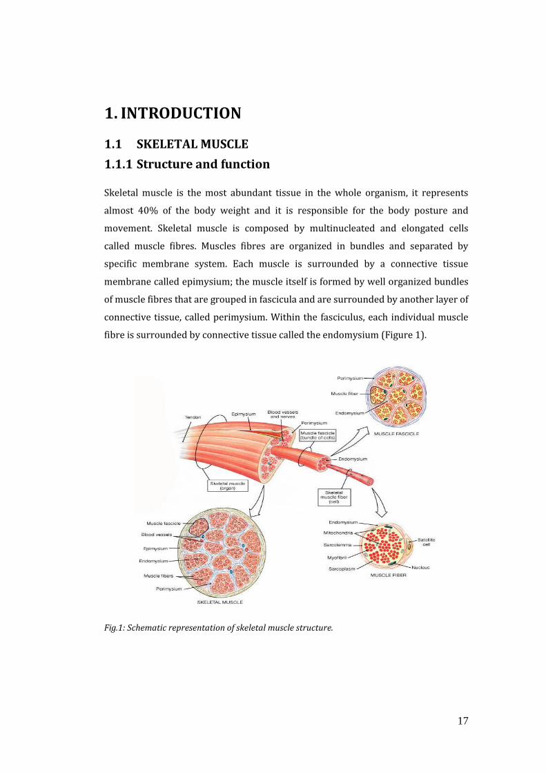
Skeletal muscle is the most abundant tissue in the whole organism, it represents
almost 40% of the body weight and it is responsible for the body posture and
movement. Skeletal muscle is composed by multinucleated and elongated cells
called muscle fibres. Muscles fibres are organized in bundles and separated by
specific membrane system. Each muscle is surrounded by a connective tissue
membrane called epimysium; the muscle itself is formed by well organized bundles
of muscle fibres that are grouped in fascicula and are surrounded by another layer of
connective tissue, called perimysium. Within the fasciculus, each individual muscle
fibre is surrounded by connective tissue called the endomysium (Figure 1).
Fig.1: Schematic representation of skeletal muscle structure.
18
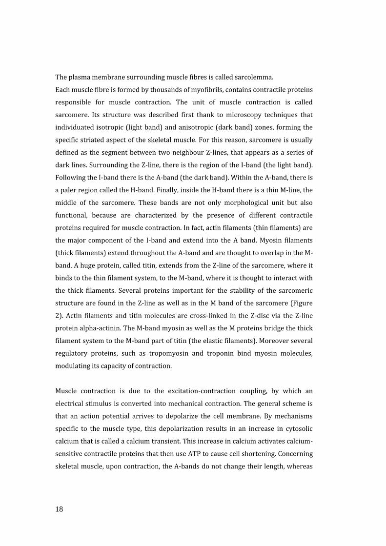
The plasma membrane surrounding muscle fibres is called sarcolemma.
Each muscle fibre is formed by thousands of myofibrils, contains contractile proteins
responsible for muscle contraction. The unit of muscle contraction is called
sarcomere. Its structure was described first thank to microscopy techniques that
individuated isotropic (light band) and anisotropic (dark band) zones, forming the
specific striated aspect of the skeletal muscle. For this reason, sarcomere is usually
defined as the segment between two neighbour Z-lines, that appears as a series of
dark lines. Surrounding the Z-line, there is the region of the I-band (the light band).
Following the I-band there is the A-band (the dark band). Within the A-band, there is
a paler region called the H-band. Finally, inside the H-band there is a thin M-line, the
middle of the sarcomere. These bands are not only morphological unit but also
functional, because are characterized by the presence of different contractile
proteins required for muscle contraction. In fact, actin filaments (thin filaments) are
the major component of the I-band and extend into the A band. Myosin filaments
(thick filaments) extend throughout the A-band and are thought to overlap in the M-
band. A huge protein, called titin, extends from the Z-line of the sarcomere, where it
binds to the thin filament system, to the M-band, where it is thought to interact with
the thick filaments. Several proteins important for the stability of the sarcomeric
structure are found in the Z-line as well as in the M band of the sarcomere (Figure
2). Actin filaments and titin molecules are cross-linked in the Z-disc via the Z-line
protein alpha-actinin. The M-band myosin as well as the M proteins bridge the thick
filament system to the M-band part of titin (the elastic filaments). Moreover several
regulatory proteins, such as tropomyosin and troponin bind myosin molecules,
modulating its capacity of contraction.
Muscle contraction is due to the excitation-contraction coupling, by which an
electrical stimulus is converted into mechanical contraction. The general scheme is
that an action potential arrives to depolarize the cell membrane. By mechanisms
specific to the muscle type, this depolarization results in an increase in cytosolic
calcium that is called a calcium transient. This increase in calcium activates calcium-
sensitive contractile proteins that then use ATP to cause cell shortening. Concerning
skeletal muscle, upon contraction, the A-bands do not change their length, whereas
19
the I bands and the H-zone shorten. This is called the sliding filament hypothesis,
which is now widely accepted. There are projections from the thick filaments, called
cross-bridges which contain the part (head) of myosin linked to actin. Myosin head
is able to hydrolyze ATP and convering chemical energy into mechanical energy. The
cross bridges are mostly oriented transverse to the fibre axis in relaxed fibres, while
angled at around 45 degrees in rigor.
Fig.2: Schematic representation of skeletal muscle sarcomere.
To allow the simultaneous contraction of all sarcomeres, the sarcolemma penetrates
into the cytoplasm of the muscle cell between myofibrils, forming membranous
tubules called transverse tubules (T-tubules) (Figure 3). The T-tubules are
electrically coupled with the terminal cisternae which continue into the
sarcoplasmic reticulum. Thus the Sarcoplasmic Reticulum, which is the enlargement
of smooth Endoplasmic Reticulum (ER) and which contains the majority of calcium
ions required for contraction, extends from both sides of T-tubules into the
myofibrils. Anatomically, the structure formed by T-tubules surrounded by two
smooth ER cisternae is called the triad and it allows the transmission of membrane
depolarization from the sarcolemma to the ER. The contraction starts when an
action potential diffuses from the motor neuron to the sarcolemma and then it
travels along T-tubules until it reaches the sarcoplasmic reticulum. Here the action

20
potential changes the permeability of the sarcoplasmic reticulum, allowing the flow
of calcium ions into the cytosol between the myofibrils. The release of calcium ions
induces the myosin heads to interact with the actin, allowing the muscle contraction.
The contraction process is ATP dependent. The energy is provided by mitochondria
which are located closed to Z line.
Fig.3: Schematic representation of organization of T-tubuls in skeletal muscle.
The contraction properties of a muscle depend on the fibre type composition.
Mammalian muscle fibres are divided into two distinct classes: type I, also called
slow fibres, and type II, called fast fibres. This classification considers only the
mechanical properties. However the different fibre types also show peculiar features
such as for example myosin ATPase enzymes, metabolism (oxidative or glycolitic),
mitochondrial content revealed by succinate dehydrogenase (SDH) staining, and
resistance to fatigue (Pette and Heilmann, 1979; Schiaffino et al., 2007). Each muscle
is composed by a combination of fibre types, whose abundance affects the type of
contraction the muscle undergoes (solw or fast); regarding that a muscle is defined
as slow, if it containing more type I fibres, or fast, if type II fibres are more abundant.
The different fibre types are also characterized by peculiar Myosin Heavy Chain
(MHC) proteins expression. The fibre type I expresses the slow isoform of MHC
(MHCβ or MHC1), and shows a great content of mitochondria, high levels of
myoglobin, high capillary densities and high oxidative capacity. Muscles containing
21
many type I fibres display red colour for the great vascularisation and for the high
myoglobin content. The type II, fast, myofibers are divided in three groups
depending on which myosin is expressed. In fact distinct genes encode for MHC IIa,
IIx (also called IId) and IIb. Type IIa myofibers are faster than type I, but they are
still relatively fatigue-resistant. IIa fibers are relatively slower than IIx and IIb and
have an oxidative metabolism due to the rich content of mitochondria (Schiaffino
and Reggiani, 1996). Given all these characteristics, IIa fibres are also termed fast
oxidative fibres. They exhibit fast contraction, high oxidative capacity and a relative
fatigue resistance. The IIx and IIb fibre types are called fast-glycolitic fibres and
show a prominent glycolitic metabolism containing few mitochondria of small size,
high myosin ATPase activity, expression of MHC IIb and MHC IIx proteins, the fastest
rate of contraction and the highest level of fatigability.
The fibre-type profile of different muscles is initially established during
development independently of neural influence, but nerve activity has a major role
in the maintenance and modulation of its properties in adult muscle. Indeed during
postnatal development and regeneration, a default nerve activity-independent
pathway of muscle fibre differentiation, which is controlled by thyroid hormone,
leads to the activation of a fast gene program. On the contrary, the post natal
induction and maintenance of the slow gene program is dependent on slow
motoneuron activity. The muscle fibre-type then undergoes further changes during
postnatal life, for example fibre-type switching could be induced in adult skeletal
muscles by changes in nerve activity (Murgia et al., 2000).
1.1.2 The Nerve-Muscle connection
Neuromuscular Junction (NMJ) development and maintenance The vertebrate skeletal neuromuscular junction (NMJ) is the connection between
motor neurons and skeletal muscle.
NMJ is the most used model in the study of single synapse, because it has a clearly
defined organization and it is quite accessible thanks to its relatively large size and
localization. NMJ lies outside the brain and post-synaptic muscle fibre is generally
innervated by one axon. Muscles are readily re-innervated following nerve damage,
22
allowing synaptogenesis to be studied also in adult and not only in embryonic
organisms. Moreover, the bond between bungarotoxins (BGT) and AChR is able to
clearly identify the localization and the morphology of NMJ.
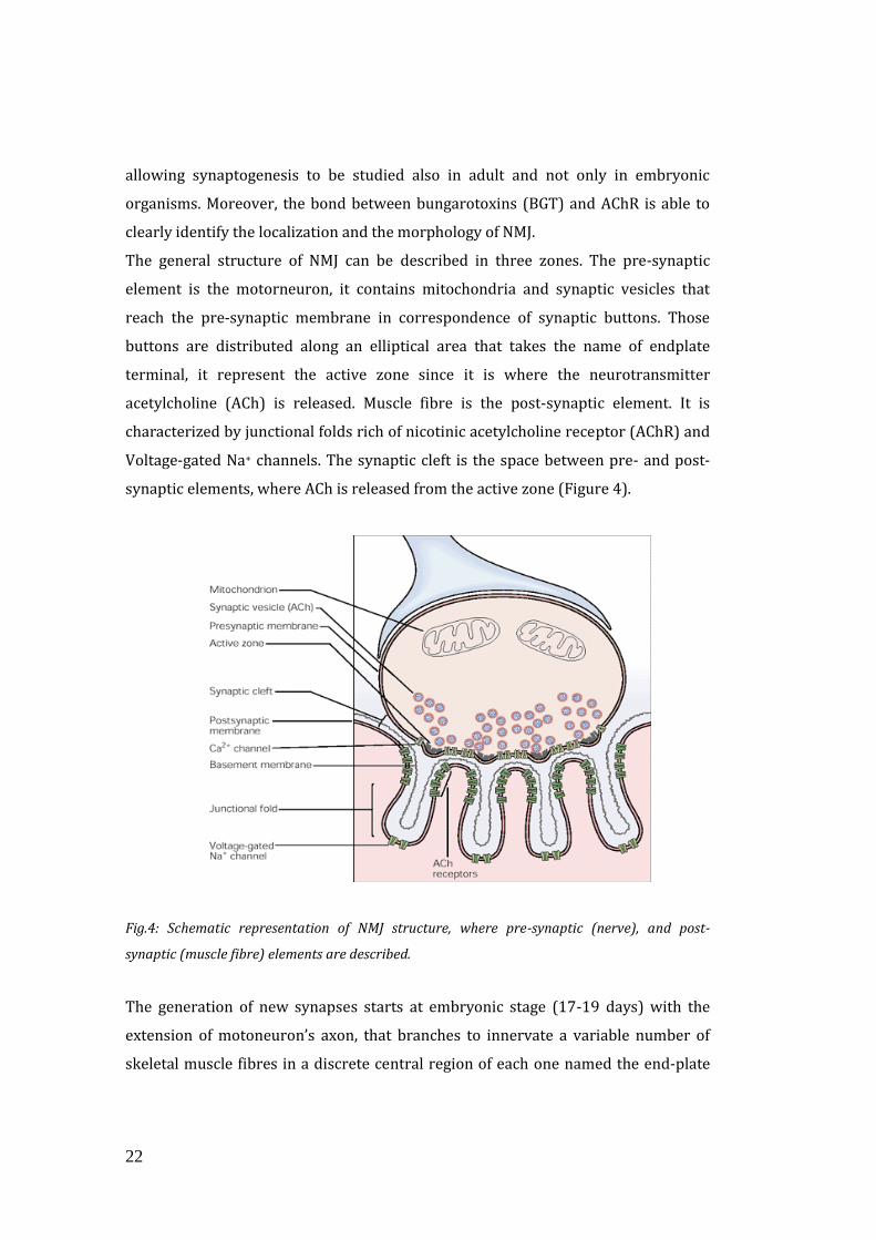
The general structure of NMJ can be described in three zones. The pre-synaptic
element is the motorneuron, it contains mitochondria and synaptic vesicles that
reach the pre-synaptic membrane in correspondence of synaptic buttons. Those
buttons are distributed along an elliptical area that takes the name of endplate
terminal, it represent the active zone since it is where the neurotransmitter
acetylcholine (ACh) is released. Muscle fibre is the post-synaptic element. It is
characterized by junctional folds rich of nicotinic acetylcholine receptor (AChR) and
Voltage-gated Na+ channels. The synaptic cleft is the space between pre- and post-
synaptic elements, where ACh is released from the active zone (Figure 4).
Fig.4: Schematic representation of NMJ structure, where pre-synaptic (nerve), and post-
synaptic (muscle fibre) elements are described.
The generation of new synapses starts at embryonic stage (17-19 days) with the
extension of motoneuron’s axon, that branches to innervate a variable number of
skeletal muscle fibres in a discrete central region of each one named the end-plate

23
band. The nerve terminal accumulates synaptic acetylcholine vesicles and other pre-
synaptic components, and while both pre- and post- synaptic membranes thicken,
the synaptic cleft widens. With increasing number of vesicles, the active zone starts
to appear and, the expression of several genes, coding for postsynaptic proteins,
including the acetylcholine receptors (AChRs), increases in the postsynaptic nuclei.
The last step of this process is axon myelination and junctional fold formation in the
post-synaptic element (Figure 5). These morphological changes in the nerve
terminal are accompanied by increased frequency of spontaneous synaptic
potential.
Fig.5: Morphological changes in NMJ development A) Representation of NMJ developmental
stages, in details (a-b) is reported the nuclei transition from extra-synaptic to the synaptic
region, where a specific transcriptional programme is activated. B) Morphological changes in
NMJ that from embrionic-oval stage became pretzel-shaped in the adult and mature one (Shi et
al., 2012).
The development of neuromuscular junction requires a fundamental process called
muscle pre-patterning that is characterized by both muscle- and nerve-specific
24
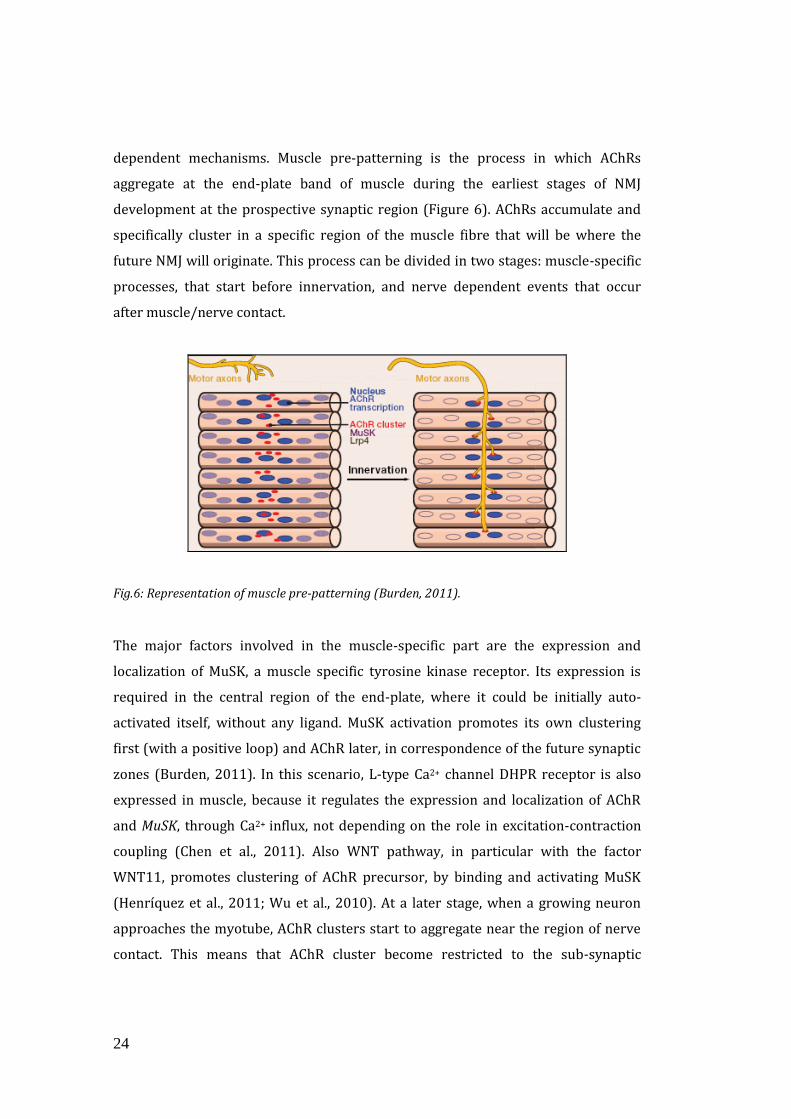
dependent mechanisms. Muscle pre-patterning is the process in which AChRs
aggregate at the end-plate band of muscle during the earliest stages of NMJ
development at the prospective synaptic region (Figure 6). AChRs accumulate and
specifically cluster in a specific region of the muscle fibre that will be where the
future NMJ will originate. This process can be divided in two stages: muscle-specific
processes, that start before innervation, and nerve dependent events that occur
after muscle/nerve contact.
Fig.6: Representation of muscle pre-patterning (Burden, 2011).
The major factors involved in the muscle-specific part are the expression and
localization of MuSK, a muscle specific tyrosine kinase receptor. Its expression is
required in the central region of the end-plate, where it could be initially auto-
activated itself, without any ligand. MuSK activation promotes its own clustering
first (with a positive loop) and AChR later, in correspondence of the future synaptic
zones (Burden, 2011). In this scenario, L-type Ca2+ channel DHPR receptor is also
expressed in muscle, because it regulates the expression and localization of AChR
and MuSK, through Ca2+ influx, not depending on the role in excitation-contraction
coupling (Chen et al., 2011). Also WNT pathway, in particular with the factor
WNT11, promotes clustering of AChR precursor, by binding and activating MuSK
(Henríquez et al., 2011; Wu et al., 2010). At a later stage, when a growing neuron
approaches the myotube, AChR clusters start to aggregate near the region of nerve
contact. This means that AChR cluster become restricted to the sub-synaptic
25
membrane, thus disappearing from the extra-synaptic one, and that AChR subunits
composition change from α2, β, δ, γ to α2, β, δ, ε resulting in acquisition of new
channel properties (Hall and Sanes, 1993; Numberger et al., 1991). AChR-ɣ subunit
is in fact a defined marker of reinnervation, because it is typical of early
developmental stages of NMJ. So when the nerve establishes contact with a muscle
cell, it exerts complex control over both number and distribution of AChRs.
There is another way in which nerve controls post-synaptic differentiation, in fact
the system that regulates the density of extra-synaptic AChR is based on electrical
activity evoked by synaptic transmission. When the nerve depolarizes the muscle,
the action potential represses AChR gene transcription in extra synaptic nuclei,
probably through Ca2+, protein kinase C and MYOD transcription factor, thus having
AChR expression specifically localized at synaptic zones. In addition, nerve terminal
can also contribute with secretion of other factors that can act locally via different
receptors (Wu et al., 2010; Hall and Sanes, 1993).
After the pre-patterning, post-synaptic differentiation takes place depending on the
clustering of neurotransmitter (NT) receptors and scaffolding proteins. These
changes are regulated by different signals including WNT pathway, and
neurotrophins secreted from the nerve. An important role is played by a molecule
called neuregulin that is secreted from the neuron, and binds muscle receptor ErbB
to induce the activation of synaptic genes (Burden, 2002; Trinidad et al., 2000). The
most important and studied signalling pathway involved in post-synaptic
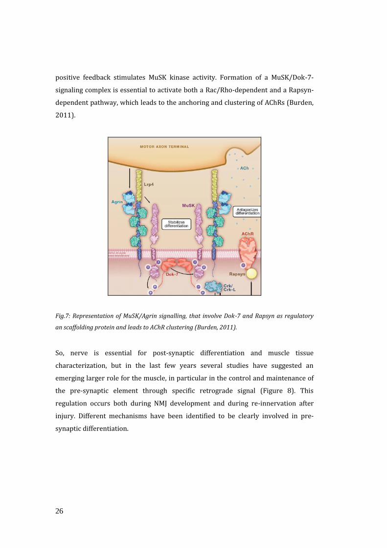
differentiation is the MuSK/Agrin signalling (Figure 7). MuSK, as already said, is a
tyrosine kinase receptor expressed in the postsynaptic membrane of NMJ, where it
co-localizes with AChRs, inducing their clustering (Kim and Burden, 2008). MuSK is
activated by Agrin, a proteoglycan released from the nerve, that stimulate MuSK
phosporylation, by interacting with Lrp4 and not directly with MuSK. Lrp4 is a
lipoprotein receptor related protein 4, and acts as co-receptor, for the Agrin-MuSK
signal. Lrp4 self-associates and interacts with MuSK also in the absence of Agrin.
Binding of Agrin to Lrp4 stimulates association between Lrp4 and MuSK and
increases MuSK kinase activity. Then MuSK stimulates recruitment of Dok-7 that is
cytoplasmic adaptor protein expressed specifically in muscle. MuSK promotes Dok-7
tyrosine phosphorylation, and after that Dok-7 is able to form a dimer and with a
26
positive feedback stimulates MuSK kinase activity. Formation of a MuSK/Dok-7-
signaling complex is essential to activate both a Rac/Rho-dependent and a Rapsyn-
dependent pathway, which leads to the anchoring and clustering of AChRs (Burden,
2011).
Fig.7: Representation of MuSK/Agrin signalling, that involve Dok-7 and Rapsyn as regulatory
an scaffolding protein and leads to AChR clustering (Burden, 2011).
So, nerve is essential for post-synaptic differentiation and muscle tissue
characterization, but in the last few years several studies have suggested an
emerging larger role for the muscle, in particular in the control and maintenance of
the pre-synaptic element through specific retrograde signal (Figure 8). This
regulation occurs both during NMJ development and during re-innervation after
injury. Different mechanisms have been identified to be clearly involved in pre-
synaptic differentiation.
27
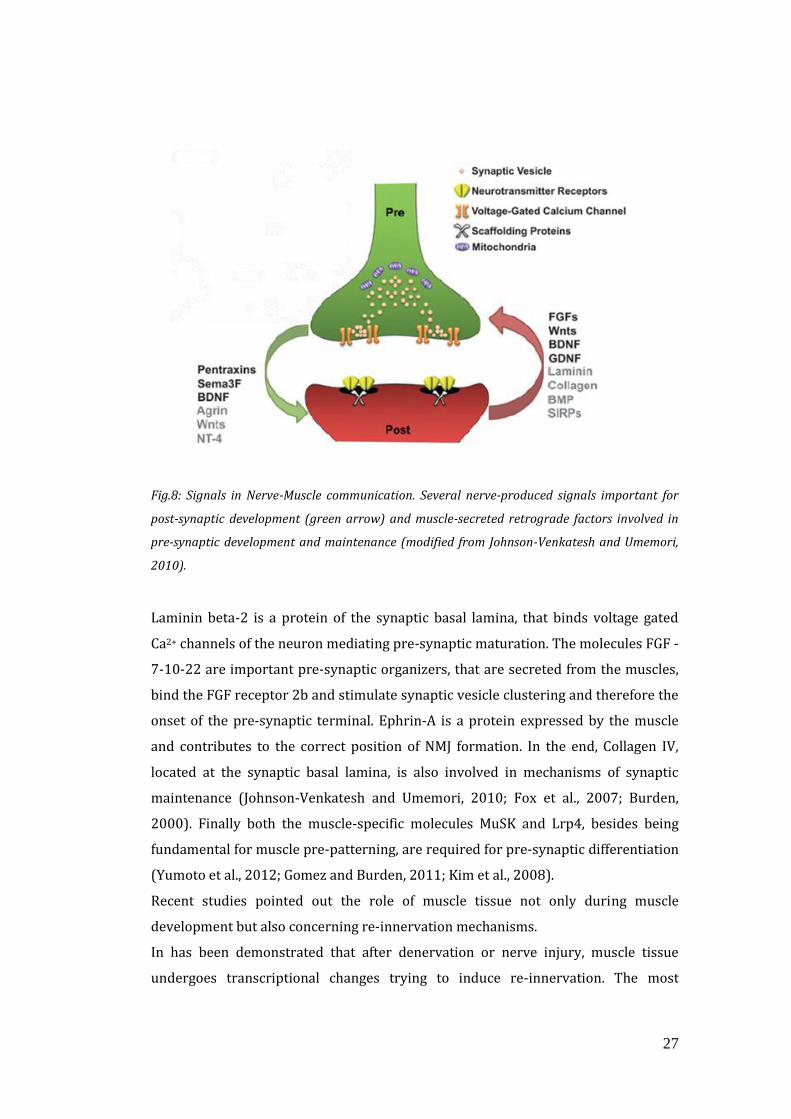
Fig.8: Signals in Nerve-Muscle communication. Several nerve-produced signals important for
post-synaptic development (green arrow) and muscle-secreted retrograde factors involved in
pre-synaptic development and maintenance (modified from Johnson-Venkatesh and Umemori,
2010).
Laminin beta-2 is a protein of the synaptic basal lamina, that binds voltage gated
Ca2+ channels of the neuron mediating pre-synaptic maturation. The molecules FGF -
7-10-22 are important pre-synaptic organizers, that are secreted from the muscles,
bind the FGF receptor 2b and stimulate synaptic vesicle clustering and therefore the
onset of the pre-synaptic terminal. Ephrin-A is a protein expressed by the muscle
and contributes to the correct position of NMJ formation. In the end, Collagen IV,
located at the synaptic basal lamina, is also involved in mechanisms of synaptic
maintenance (Johnson-Venkatesh and Umemori, 2010; Fox et al., 2007; Burden,
2000). Finally both the muscle-specific molecules MuSK and Lrp4, besides being
fundamental for muscle pre-patterning, are required for pre-synaptic differentiation
(Yumoto et al., 2012; Gomez and Burden, 2011; Kim et al., 2008).
Recent studies pointed out the role of muscle tissue not only during muscle
development but also concerning re-innervation mechanisms.
In has been demonstrated that after denervation or nerve injury, muscle tissue
undergoes transcriptional changes trying to induce re-innervation. The most
28
important defined signals are the over-expression of MuSK and AChR-ɣ subunit and
also miR206 that promotes the expression of FGFBP1, leading to muscle re-
innervation (Williams et al., 2009). FGFBP1 is a secreted protein that interacts and
potentiates the bioactivity of FGF-7, FGF-10, and FGF-22 family members (Jang and
Van Remmen, 2011; Williams et al., 2009).
At present the characterization of the mechanisms of muscle-nerve communication
is an open issue.
1.2 MUSCLE HYPERTROPHY AND ATROPHY
Skeletal muscle mass is orchestrated by several complex mechanisms that regulate
the rate of muscle growth and muscle loss. Muscle growth is mainly due to protein
synthesis, that, when exceeds, leads to muscle hypertrophy. On the contrary,
excessive protein degradation, loss of organelles and cytoplasm are major causes of
muscle atrophy.
Muscle Hypertrophy
Skeletal muscle hypertrophy is defined as an increase in muscle mass, which in the
adult animal comes as a result of an increase in the size of pre-existing skeletal
muscle fibres. This growth, as stated above, is mainly due to increased protein
synthesis and concomitant decreased protein degradation. The Insulin-like growth
factor (IGF-1)-AKT signalling is the major pathway that controls muscle growth.
Muscle-specific IGF-1 over-expression in transgenic mice results in muscle
hypertrophy and, importantly, the growth of muscle mass matches with a
physiological increase of muscle strength. Furthermore, the over-expression of a
constitutively active form of AKT, a downstream target of IGF-1, in adult skeletal
muscle induced muscle hypertrophy. Moreover, AKT transgenic mice display muscle
hypertrophy and protection from denervation-induced atrophy, showing that AKT
pathway promotes muscle growth and simultaneously blocks protein degradation
(Schiaffino et al., 2013). AKT pathway, in fact, controls in an opposite manner two
29
important downstream targets: mammalian target of rapamycin (mTOR) and
glycogen synthase kinase 3 beta (GSK3β). In the first case, AKT activates mTOR, that
is a key regulator of cell growth, promoting the activation of S6 kinase (S6K) and
blocking the inhibition of eif4e binding protein 1 (4EBP1) on eukaryotic translation
initiation factor 4E (eif4e), thus leading to protein synthesis. In the other case, the
inhibition of GSK3β from AKT stimulates proteins synthesis, since GSK3β normally
blocks protein translation initiated by eIF2B protein (Glass, 2005). Taken together
with other observations, these results suggest that IGF-1- AKT axis is a major
mediator of skeletal muscle hypertrophy. Recently, it has been reported that also
TGF-β pathway contributes to regulation of muscle mass in adulthood (Sartori et al.,
2013). Sartori et al. showed that when the BMP pathway is blocked or myostatin
expression is increased, more Smad4 is available for phosphorylated Smad2/3,
leading to an atrophy response. Therefore, under normal circumstances, a balance
between these competing pathways is required to maintain muscle mass. Moreover
they identify a newly characterized ubiquitin-ligase, named MUSA1, as the molecular
mechanism underlying the anti-atrophic action of the BMP pathway that has a
negative effect on its expression. This work provided evidences that also BMP
signalling is involved in the regulation of adult muscle mass in normal and
pathological situations (Sartori et al., 2013).
Muscle atrophy
Atrophy is defined as a decrease in cell size mainly due to protein degradation and
then to the loss of organelles and cytoplasm as well. This is because protein turnover
is dominant over cellular one during acute phases of muscle wasting, for example
when sarcomeric proteins are rapidly lost during fasting, disuse, and denervation.
Muscle loss is mediated by two highly conserved pathways: ubiquitin-proteasomal
system (UPS) and autophagy-lisosomal pathway (ALP).
Several evidences strongly support a major role of UPS during muscle loss. In this
process Ubiquitin (Ub) is covalently attached to substrate proteins via a three-step
mechanism involving the sequential actions of E1 (ubiquitin-activating enzyme), E2
(ubiquitin-conjugating enzyme) and E3 (ubiquitin ligase) enzymes. The rate limiting
30
enzyme of UPS is the E3 which catalyzes the transfer of ubiquitin from the E2 to the
lysine in the substrate. This reaction is highly specific and the proteins, committed to
ubiquitination and to proteasomal degradation, are recognized by the E3. Thus the
amount and the type of proteins degraded by the proteasome is largely determined
by which E3 ligases are activated in the cell (Gomes et al., 2001).
FoxO3 transcription is the key regulator of these systems, being it necessary and
sufficient for the induction of autophagy in skeletal muscle in vivo (Mammucari et al.,
2007; Zhao et al., 2007). Moreover it induces the transcription of two fundamental
muscles ubiquitin-ligases: Atrogin-1 and MuRF-1 (Sandri et al., 2004; Gomes et al.,
2001; Bodine, 2001). These ubiquitin-ligases were identified through gene
expression profile analysis performed on different atrophic models, as part of a set
of genes, called ‘atrogenes’, that triggered or were involved in the atrophic program.
These genes encode for proteins involved in different cellular processes like energy
production, transcription factors, regulators or protein synthesis and enzymes of
metabolic pathways. Among the upregulated atrophy-related genes there is a subset
of transcripts related to protein degradation pathways.
Together these findings indicate that muscle atrophy is a process that requires the
activation of a specific transcriptional program.
1.3 AGEING IN MUSCLE TISSUE: SARCOPENIA
During ageing muscle undergoes an inevitable loss of muscle mass accompanied by
loss of force, that is called sarcopenia. It has been estimated that 25% of people
under the age of 70, and 40% of people aged 80 or older are sarcopenic. As people
age, the strength in their muscles gradually decreases at a rate of 1-2% per year
after the age of 50 and by 30-40% at the age of 70 (Rossi et al. 2008). This condition
profoundly contributes to a reduced quality of life in elderly and predisposes them
to an increased risk of morbidity, disability and mortality (Visser and Schaap, 2011).
Despite the clinical, social and economic relevance of sarcopenia, the precise
mechanisms for the age-related loss of muscle mass and function are not yet fully
understood. Age-related changes in muscle are complex with key features including
myofibre atrophy, profound weakness that is partially independent from muscle
31
mass loss, myofibre degeneration, accumulation of dysfunctional mitochondria and
increased oxidative stress.
Until recently, it was thought that age-associated atrophy and weakness were
secondary to motor-neuron loss in the brain or in spinal cord. However this
hypothesis has been recently challenged. In fact little neuronal death occurs in most
areas of ageing nervous system and there is no decline of lower motor neurons
during ageing (Chai et al., 2011; Morrison and Hof, 1997). Conversely, it is emerging
that the neuromuscular junctions (NMJ) and their interactions with myofibres are
greatly altered during ageing, resulting in a loss of muscle innervation (Chai et al.,
2011; Valdez et al., 2010). In particular, loss of nerve endings has been reported at
motor endplates in both rodents and humans, indeed in the soleus muscle of aged
(22-month old) rats, both pre- and post-synaptic specializations were significantly
smaller compared to that of the young (8-month old) rats (Deschenes and Wilson,
2003). In this way, the muscle fibre seems to have an important role in the
degeneration of motoneuron. It has been shown that reorganization of AChR plaque
into multiple fragments was an occasional event that followed the degeneration of
the underlying muscle fibre. Moreover, the increased prevalence of fragmented
endplates in elderly was attributed to an increased incidence of sporadic muscle
fibre degeneration events as the animal grew older (Li et al., 2011).
Skeletal muscle tissue is particularly vulnerable to oxidative stress, in fact being a
post mitotic tissue, it uses large amount of oxygen, thus causing cumulative
oxidative damage to the cell structures over time. Sarcopenic muscle degeneration is
associated to an age-related oxidative stress that leads to increased mitochondrial
DNA damage, lipid peroxidation and protein oxidation. A great number of studies
have shown an increase in oxidized proteins at the intracellular level during
senescence, this causes loss of function in the affected protein that could lead to
their accumulation, compromising organ functionality (Rossi et al., 2008). Oxidative
stress and decreased release of trophic factors are considered two independent and
important causes that affect NMJ integrity and contribute to denervation, also
during ageing process (Jang and Van Remmen, 2011). Several laboratories have
32
tested the impact of oxidative stress on age-related muscle wasting. In order to
elucidate the direct cause and effect relation between oxidative stress and
sarcopenia in vivo, a mouse holding homozygous deletion of an essential antioxidant
enzyme Sod1 (Cu/Zn superoxide dismutase, Cu/Zn SOD) was generated (Jang et al.,
2010). It has been shown that the lack of Sod1 led to age-dependent muscle atrophy
with alterations in NMJs, that were similar to normal ageing muscle but occurred
earlier and more frequently (Jang et al., 2010). These data indicated that
maintenance of NMJ during ageing may be critically influenced by oxidative stress.
For this reason it has been investigated whether mitochondrial dysfunction in the
population of mitochondria associated with the NMJ may lead to altered calcium
buffering and oxidative modification of key molecules in the NMJ, thus contributing
to age-associated declines in neuromuscular innervation. Zhou et al. demonstrated
that mitochondria adjacent to the AChR are selectively depolarized when muscle
fibres are challenged by calcium in a mouse model of ALS, which exhibits significant
neuromuscular degeneration (Zhou et al., 2010). In addition, other works have
shown that muscle-specific over-expression of uncoupling proteins (UCP1)
significantly disrupted NMJ integrity. Furthermore, it has been previously reported
that isolated subsarcolemmal mitochondria of Sod1-/- mice have significant deficits
in ATP generation and oxygen consumption, and also generate more mitochondrial
ROS compared to wild-type (Jang et al., 2010).
The exchange of trophic factors is implicated in pre- and post- synaptic development
as well as in the preservation of neuronal and synaptic plasticity at the NMJ. The
exact role or the identity of neurotrophic and/or myotrophic factors that promote
survival and maintenance of pre-synaptic and post-synaptic apparatus at the NMJ, in
the context of ageing, has not been fully determined. However, recent studies
indicate that a variety of trophic factors such as brain derived neurotrophic factor
(BDNF), neutrophin-3 (NT-3), neutrophin-4 (NT-4), cytokines such as glial-derived
neutrophic factor (GDNF) and ciliary neutrophin factor (CNTF), and other growth
factors as insulin-like growth factor (IGF-1 and IGF-II) and fibroblast growth factors
(FGF), play a modulatory role in neuromuscular system to a different extent during
ageing (Jang and Van Remmen, 2011).

33
Up to now it is not known whether myofibre denervation is due to deleterious
changes, such as impaired trophic factors production or increased oxidative stress,
in muscle cells themselves, in neurons or both components.
Notably, two lifestyle adaptations, namely caloric restriction and exercise, have been
consistently demonstrated to extend lifespan and, in parallel, to mitigate age-related
alterations of NMJ (Melov et al., 2007; Fontana et al., 2010; Sandri et al., 2013;
Schiaffino et al., 2013; Coen et al., 2013; Toledo et al., 2013; Guarente, 2013).
It has been shown that caloric restriction directly attenuates age-related loss of
muscle mass by improving mitochondria function, which in turn, lowers the
mitochondrial ROS production in Sod-/- mice. Those effects of caloric restriction on
mitochondria contribute to the preservation of NMJ morphology, innervation of
muscle fibres, and maintenance of muscle mass and structure, improving also the
regenerative potential of skeletal muscle, that normally decrease with age (Jang et
al., 2012).
Physical activity is known to trigger several changing in the muscle tissue such as
switching of fibre type, increase in mitochondrial biogenesis, metabolic variation in
glucose consumption and lactate production, activation of AMPK. AMPK is the AMP-
activated protein kinase that plays an important role in cellular energy homeostasis.
In fact it is involved in many different pathways and is considered the master sensor
of energy imbalance, as the major regulator of metabolic switch upon stress
condition. When activated, it phosphorylates its direct target, acetyl-CoA carboxilase
(ACC) and contributes to translocation of the glucose transporter Glut 4 on the cell
surface (Kurth-Kraczek et al., 1999). Glut 4 is a muscle specific isoform of glucose
transporter, and it is normally located in intracellular storage sites, and move to the
cell surface in response to insulin, muscle contraction, and other stimuli that
requires an increased glucose transport (Holloszy, 2011).
Physical activity also triggers some beneficial effects on NMJ maintenance. Exercise
in fact could partially reverse NMJ structural alterations that had already occurred
after a denervation event (Valdez et al., 2010). Moreover since beneficial effects
were observed in exercised muscles only, the ameliorated phenotype of the synapse
resulted from local muscle-nerve interactions, suggesting that increased activity in
34
exercising muscles could lead to an up-regulation of trophic factors from muscle that
would, in turn, improve synaptic maintenance (Valdez et al., 2010). These findings
correlate with another work by Cheng (Cheng et al., 2013), where 21 months old
mice and 18 months old mice, with reduced nerve terminal size, performed
respectively 4 and 10 months voluntary wheel running. The authors found that after
exercise most of the age-associated loss of nerve terminal area was prevented
(Cheng et al., 2013). Furthermore, the positive effects of exercise are not limited to
NMJ, but can be extended to a more general action to prevent ageing. In fact, 5
months of exercise training were sufficient to completely reverse the premature
ageing phenotype of the mitochondrial DNA mutator mice, which possess a
dysfunctional copy of the mitochondrial proofreading-exonuclease, polymerase
gamma (Safdar et al., 2011).
It is important to consider another aspect that occurs during physical activity:
contractions produce free radicals and ROS production is potentially damaging to
the muscle tissues (Powers and Jackson, 2008).
Several studies tried to identify ROS sources during exercise and a number of
researchers have assumed that the increased ROS generation that occurs during
contractile activity is directly related to the elevated oxygen consumption that
occurs with increased mitochondrial activity (Kanter et al., 1994; Urso et al., 2003).
Although mitochondria are involved in ROS production upon exercise, other studies
pointed out that they are not the only source of ROS in skeletal muscle during
exercise. Several works found NADH-oxidase enzyme associated with the
sarcoplasmic reticulum (SR) of both cardiac and skeletal muscle, and it was
responsible for the superoxide production. Thus, in this case, the superoxide
generation influenced calcium release by the SR through oxidation of the ryanodyne
receptor (Cherednichenko et al., 2004; Xia et al., 2003).
Some recent findings proposed a new essential role for exercise-induced ROS
formation, in promoting insulin sensitivity in humans, supporting the notion that
anti-oxidants are detrimental for exercise-induced benefits in humans, although the
mechanisms remain unclear (Ristow et al., 2011).
Even it has been widely investigated the role of ROS during exercise is still not
completely clear.
35
1.4 THE AUTOPHAGY-LYSOSOMAL SYSTEM
The autophagy-lysosomal pathway is an evolutionarily conserved catabolic process
essential for metabolic homeostasis maintenance, depending on nutrient
availability. This process is responsible for the degradation of cytosolic component,
long-lived proteins, damaged organelles, protein aggregates and intracellular
pathogens. Autophagy in fact takes place at basal levels in all eukaryotic cells to
maintain or rejuvenate function of proteins and organelles, but can also be induced
by limitation of various types of nutrients, such as amino acids, growth factors,
oxygen and energy as an adaptive mechanism essential for cell survival (Mizushima,
2011). During the autophagy process, the cargo that needs to be degraded is
engulfed by double membranes layer called autophagosomes. These membranes
have to be committed, and this requires the recruitment of ATGs proteins on the
membrane, as I will explain hereafter. Then the vesicles are delivered to the
lysosomes where the cargo is degraded to amino acids that supply energy
requirement. This role in recycling is complementary to that of the ubiquitin-
proteasome system, which degrades proteins to generate oligopeptides that are
subsequently degraded into amino acids (Lecker et al., 2006).
The autophagy system is highly regulated through the action of various kinases,
phosphatases, and guanosine triphosphatases (GTPases). The core protein
machinery that is necessary to commit membranes to become vesicles includes two
ubiquitin-like protein conjugation systems (Sandri, 2010). Moreover there is
another set of proteins, that regulates the vesicle formation and their docking and
fusion with lysosome (Boya et al., 2013).
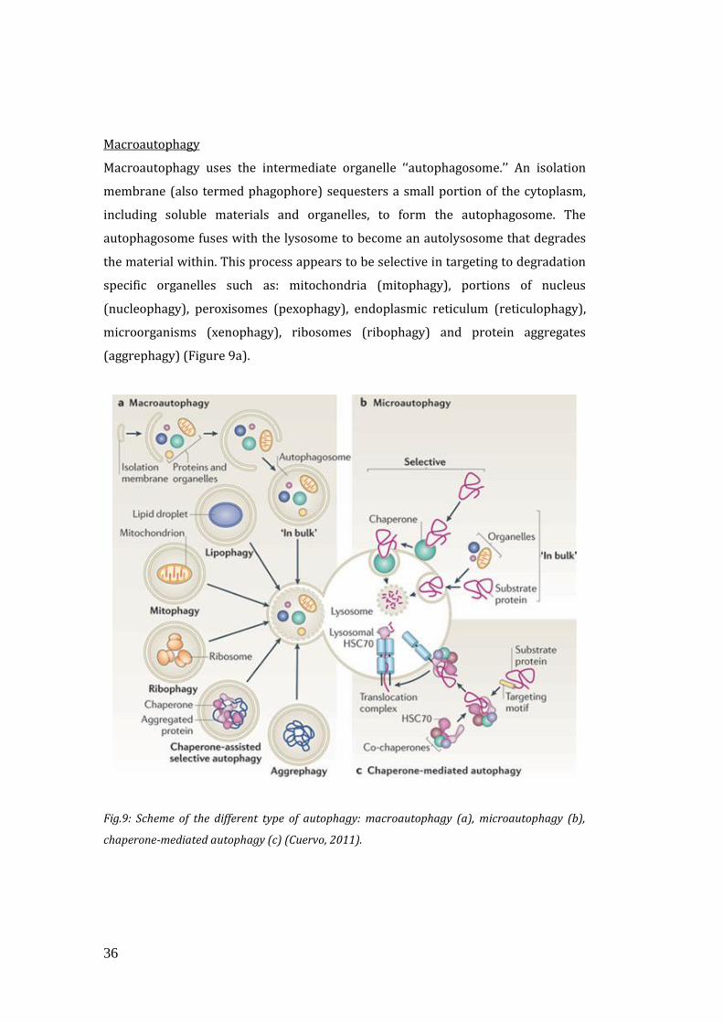
There are mainly three classes of autophagy: macroautophagy, microautophagy, and
chaperone-mediated autophagy (Figure 9).
36
Macroautophagy
Macroautophagy uses the intermediate organelle ‘‘autophagosome.’’ An isolation
membrane (also termed phagophore) sequesters a small portion of the cytoplasm,
including soluble materials and organelles, to form the autophagosome. The
autophagosome fuses with the lysosome to become an autolysosome that degrades
the material within. This process appears to be selective in targeting to degradation
specific organelles such as: mitochondria (mitophagy), portions of nucleus
(nucleophagy), peroxisomes (pexophagy), endoplasmic reticulum (reticulophagy),
microorganisms (xenophagy), ribosomes (ribophagy) and protein aggregates
(aggrephagy) (Figure 9a).
Fig.9: Scheme of the different type of autophagy: macroautophagy (a), microautophagy (b),
chaperone-mediated autophagy (c) (Cuervo, 2011).
37
Microautophagy
In microautophagy, the lysosome itself engulfs small components of the cytoplasm
by inward invagination of the lysosomal membrane. Membrane dynamics during
microautophagy may be quite similar or identical to that of endosomal sorting
complex required for transport (ESCRT)-dependent multivesicular body (MVB)
formation, which occurs in the late endosome (Figure 9b).
Chaperone-Mediated Autophagy (CMA)
The third type of autophagy is chaperone-mediated autophagy (CMA). This class
does not involve membrane reorganization; instead, substrate proteins directly
translocate across the lysosomal membrane. The chaperone protein Hsc70 (heat
shock cognate 70) and co-chaperones specifically recognize cytosolic proteins that
contain a KFERQ-like pentapeptide. The transmembrane protein Lamp-2A, which is
an isoform of Lamp-2, acts as a receptor on the lysosome, and unfolded proteins are
delivered into the lysosomal lumen through a multimeric translocation complex
(Mizushima, 2011) (Figure 9c).
Macroautophagy, hereafter called autophagy, is thought to be the major type of
autophagy, and it has been studied most extensively compared to microautophagy
and CMA.
Autophagy is activated by both caloric restriction and exercise (Grumati et al., 2010;
Grumati et al., 2011a; Grumati et al., 2011b; He et al., 2012; Rubinsztein et al., 2011;
Wohlgemuth et al., 2010).
Although many studies focused on these topic the mechanism that link autophagy to
these lifestyle conditions is still under investigation.
1.4.1 The autophagy genes
Genetic screens in S. cerevisiae have led to the identification of a number of
molecular factors essential for autophagy. There are currently over 30 genes that
are primarily involved in bulk and selective types of autophagy and they have been
named autophagy-related genes (ATG) (Klionsky et al., 2003). ATGs encode for
38
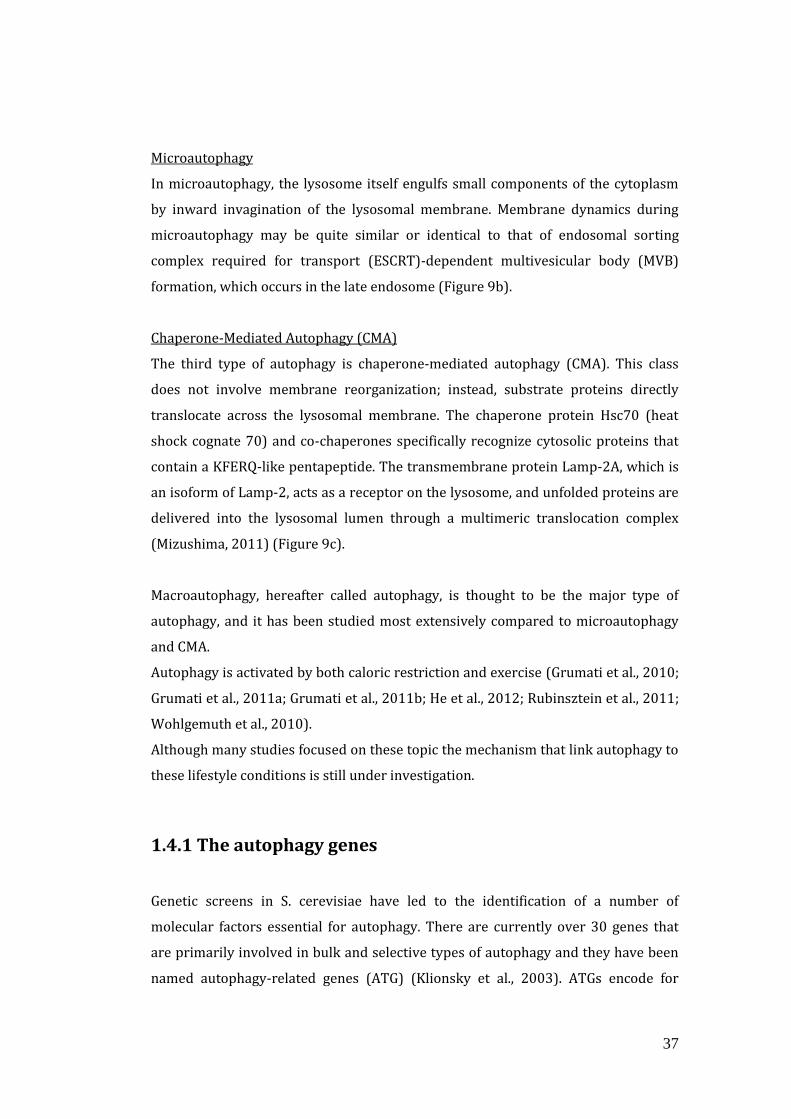
proteins that mediate the autophagic process; in particular they orchestrate the
following steps: initiation, elongation, maturation and fusion of the autophagosome
with the lysosome, and cargo degradation (Tan, 2013) (Table 1).
Table 1: ATG proteins. The 15 conserved autophagy-related gene (Atg) proteins involved in
double-membrane vesicle formation (adapted from Tan, 2013). In the left column are reported
the mammalian proteins, while on the right the homologue ones in yeast.
1.4.2 Autophagy Machinery
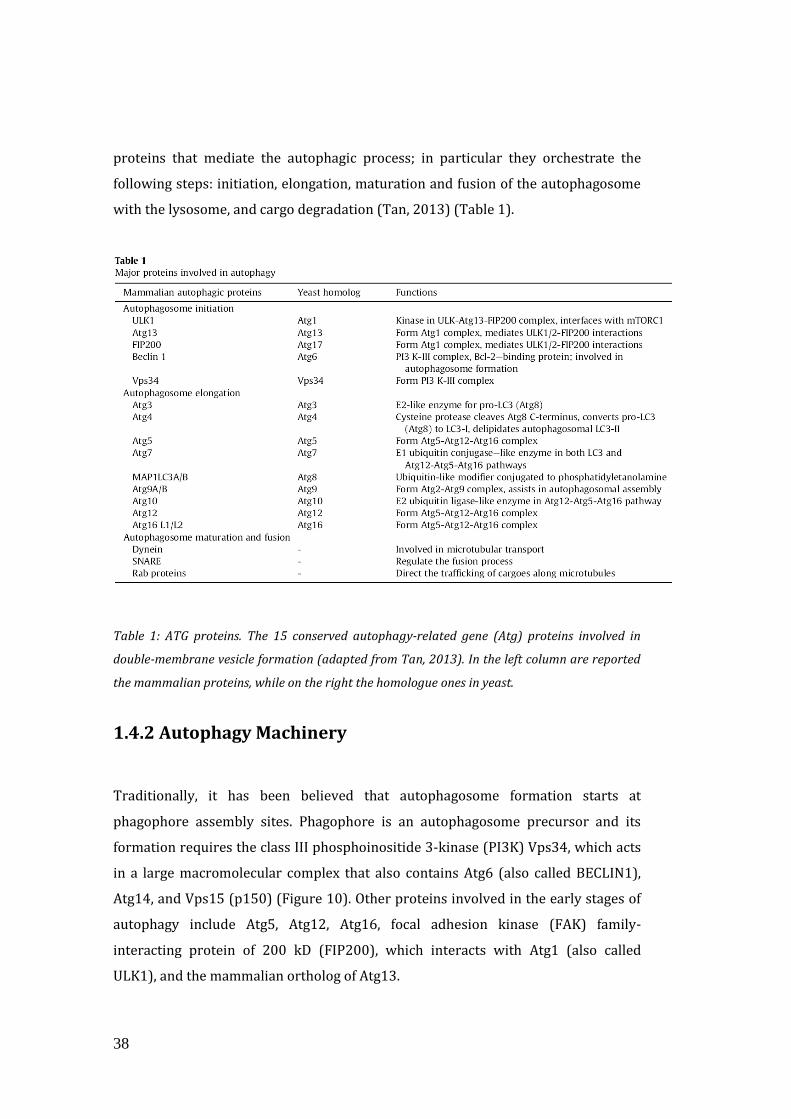
Traditionally, it has been believed that autophagosome formation starts at
phagophore assembly sites. Phagophore is an autophagosome precursor and its
formation requires the class III phosphoinositide 3-kinase (PI3K) Vps34, which acts
in a large macromolecular complex that also contains Atg6 (also called BECLIN1),
Atg14, and Vps15 (p150) (Figure 10). Other proteins involved in the early stages of
autophagy include Atg5, Atg12, Atg16, focal adhesion kinase (FAK) family-
interacting protein of 200 kD (FIP200), which interacts with Atg1 (also called
ULK1), and the mammalian ortholog of Atg13.
39
Fig.10: Possible origins for the membrane of the nascent autophagosome. This image reports
the major players involved in the autophagosome formation (Rubinsztein et al., 2011).
The elongation of the autophagosomal membranes is a critical step that is associated
with two ubiquitination-like reactions. In the first one, Atg12 is conjugated to Atg5
by Atg7, which is an E1 ubiquitin-like activating enzyme, and Atg10, which is an E2
ubiquitin-like conjugating enzyme. The Atg5-Atg12 conjugates interact non
covalently with Atg16L1 and the whole complex associates with growing
phagophores but dissociates once the autophagosomes is complete (and so the
membrane is closed). In the second ubiquitin-like reaction, microtubule-associated
protein 1 light chain 3 (MAP-LC3/Atg8/LC3) is conjugated to the lipid
phosphatidylethanolamine (PE) with a covalent bond, by Atg7 (E1-like) and Atg3
(E2- like) to form LC3-II (Figure 11) (Rubinsztein et al., 2011). This modification
results in a change of the molecular weight of LC3 that allows to distinguish by
western blot the cytoplasmic soluble LC3-I, from the lipidated LC3-II isoforms.
Usually, the accumulation of LC3-I or a decrease in LC3II means that autophagy is
blocked.

40
Fig.11: Ubiquitination-like reactions in the autophagosome formation: Atg12-Atg5 conjugation
system and LC3 lipidation process (adapted from Kroemer et al., 2010).
LC3-II is required both on the outer and inner membranes of the nascent
autophagosome. Sequestered organelles and proteins are then docked to the
lysosomes for their degradation. The fusion between the outer autophagosomal
membrane with the lysosomal one also determines the degradation of the inner
membrane and of the proteins that are associated with it. Because of the transient
nature of the autophagosomes, the lifetime of LC3 and its homologues is rather
short. This feature represents the main difference between the ubiquitin-
proteasome system and the autophagy-lysosome one, in fact the fate of the ubiquitin
and ubiquitin-like proteins is different. While the ubiquitin proteasome pathway
recycles ubiquitin molecules, the autophagy-lysosome system progressively loses
the ubiquitin-like proteins, forcing the cell to replenish them in order to maintain
the autophagic flux. Multiple LC3-positive autophagosomes form randomly in the
cytoplasm, then they are trafficked along microtubules in a dynein-dependent
manner to lysosomes, which cluster close to the microtubule organizing center
(MTOC) near the nucleus. Autophagosome-lysosome fusion appears to be mediated
by the SNARE proteins VAMP8 and Vti1B (Rubinsztein et al., 2011).
Selective autophagy relies on cargo-specific autophagy receptors that facilitate cargo
sequestration into autophagosomes. Autophagy receptors directly interact with the
structure that needs to be specifically eliminated by autophagy, as well as with the
41
pool of the Atg8 (yeast homologue of mammalian LC3) protein family members
present in the internal surface of the growing autophagosomes. The latter
interaction is mostly mediated through a specific amino acid sequence present in the
autophagy receptors and commonly referred to as the LC3-interacting region (LIR)
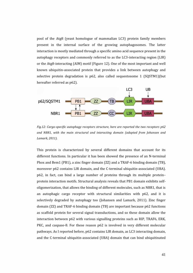
or the Atg8-interacting (AIM) motif (Figure 12). One of the most important and well
known ubiquitin-associated protein that provides a link between autophagy and
selective protein degradation is p62, also called sequestosome 1 (SQSTM1)(but
hereafter referred as p62).
Fig.12: Cargo-specific autophagy receptors structure, here are reported the two receptors p62
and NBR1, with the main structural and interacting domain (adapted from Johansen and
Lamark, 2011).
This protein is characterized by several different domains that account for its
different functions. In particular it has been showed the presence of an N-terminal
Phox and Bem1 (PB1), a zinc finger domain (ZZ) and a TRAF-6 binding domain (TB),
moreover p62 contains LIR domain, and the C-terminal ubiquitin-associated (UBA).
p62, in fact, can bind a large number of proteins through its multiple protein–
protein interaction motifs. Structural analysis reveals that PB1 domain exhibits self-
oligomerization, that allows the binding of different molecules, such as NBR1, that is
an autophagic cargo receptor with structural similarities with p62, and it is
selectively degraded by autophagy too (Johansen and Lamark, 2011). Zinc finger
domain (ZZ) and TRAF-6 binding domain (TB) are important because p62 functions
as scaffold protein for several signal transductions, and so these domain allow the
interaction between p62 with various signalling proteins such as RIP, TRAF6, ERK,
PKC, and caspase-8. For these reason p62 is involved in very different molecular
pathways. As I reported before, p62 contains LIR domain, as LC3 interacting domain,
and the C-terminal ubiquitin-associated (UBA) domain that can bind ubiquitinated
42
proteins. Recent studies have identified the LC3 recognition sequence (LRS) in
murine p62, that is located between the zinc finger and UBA domains and has the
same function of LIR in the human one. In this way p62 acts as a bridge between
ubiquitinated proteins that has to be degraded and LC3-II located in the inner
membrane of the autophagosome. Since LC3-II in the inner autophagosomal
membrane is degraded together with other cellular constituents by lysosomal
proteases, p62 trapped by LC3 is transported selectively into the autophagosome,
and the impaired autophagy is accompanied by accumulation of p62 (Ichimura and
Komatsu 2010). p62 is more linked to the autophagy-lysosome system than to the
ubiquitin-proteasome system. In fact inhibition of lysosomal degradation but not
proteasomal one, results in important accumulation of p62 (Bjørkøy et al., 2005;
Pankiv et al., 2007). Accumulation of p62 results in self-oligomerization and
formation of aggregates that contain polyubiquitinated proteins. Moreover, tissue
specific inhibition of autophagy leads to a rapid and robust increase in p62 protein
levels (Komatsu et al., 2007). There is crosstalk between different degradation
pathways for misfolded proteins, indeed the loss of one degradation system may
result in the activation of other systems. However, a high constitutive level of p62
caused by autophagy inhibition may itself contribute to an increased formation or
decreased degradation of Ub-proteins. It is suggested that p62 accumulation due to
autophagy inhibition delays the delivery of ubiquitinated proteins to the
proteasome (Johanse and Lamark, 2011). p62 is also required in the targeting to the
autophagosomes of dysfunctional mytochondria, thus being involved in the specific
selective autophagy process, called mitophagy.
1.4.3 Mitophagy
Mitochondria are crucial organelles in the production of energy and in the control of
signalling cascades. Moreover mitochondria are dynamic organelles, often organized
in the cytoplasm as a network, a reticulum of interconnected organelles shaped by
fusion (joining individual mitochondria together to become one) and fission
(dividing one mitochondrion into two mitochondria) processes. When either
process is blocked, the unopposed progression towards the other side of the
43
equilibrium defines how mitochondria appear. In mammalian cells, mitochondrial
fission depends on dynamin-related protein 1 (DRP1), and FIS1, on the contrary
fusion process depends on two mitofusins (Mfn1 and Mfn2) and the protein optic
atrophy 1 (OPA1) (Scorrano, 2013; Hall et al., 2013). Several studies demonstrated
that mitochondria shaping machinery is involved in the response of essential
changes in the cell (Romanello et al., 2010; Gomes et al., 2011).
Since mitochondria are fundamental and required for several physiological
pathways, a well defined mitochondria control and turnover is essential for cell
homeostasis. Moreover, cells must remove damaged mitochondria to prevent the
accumulation of ROS. The control of mitochondrial quality is mediated by
mitophagy, a specific type of macroautophagy, that is very important in preventing
ageing, neurodegenerative diseases, and other pathologies. In response to
potentially lethal stress or damage, mitochondrial membranes undergo
permeabilization (MMP), that constitutes one of the hallmarks of imminent
apoptotic or necrotic cell death (Kroemer et al., 2007). If only a fraction of
mitochondria is permeabilized, autophagic removal of damaged mitochondria can
rescue the cell.
Fig.13: Mitochondrial recruitment during mitophagy: (A) Atg32 in yeast, NIX and BNIP in
mammals bind mitochondria that has to be degraded, (B) moreover mitophagy can be
stimulated by mitochondrial fission through DRP1 activation (adatpted Dodson et al., 2013).
The integrity of mitochondrial membrane is essential for the maintenance of the
mitochondrial membrane potential, for this reason when the mitochondrial
44
membrane is permeabilized, mitochondria are not able to maintain the potential
anymore, becoming depolarized.
The autophagic recognition of depolarized mitochondria is mediated by a voltage
sensor, involving the mitochondrial kinase PINK1. Under normal condition, PINK1 is
continuously recruited to the mitochondrial outer membrane and degraded through
a voltage-dependent proteolysis, which leads to its removal from mitochondria first,
and to proteasome-mediated degradation later (Narendra et al., 2010). Upon
mitochondrial depolarization, PINK1 rapidly accumulates on the mitochondrial
surface, and facilitates the recruitment of the E3 ubiquitin ligase PARKIN (Narendra
et al., 2010). PARKIN ubiquitinates mitochondrial substrates including the outer
membrane protein VDAC1 and Mitofusin (Mfn), recruits the autophagy adaptor
molecule, p62/SQSTM1, and thus targets mitochondria for autophagic removal
(Geisler et al., 2010). Mitophagy can be mediated by specific factors, such as Atg32 in
yeast, NIX and BNIP in mammals, that target to mitochondria and bind to LC3
(Figure 13A); it can also be stimulated by mitochondrial fission through DRP1
activation. (Dodson et al., 2013; Lee et al., 2012)(Figure 13B).
1.4.4 Molecular signalling in autophagy
Autophagy is induced by a variety of stress stimuli, including nutrient and energy
stress, ER stress, pathogen-associated molecular patterns (PAMPs) and danger-
associated molecular patterns (DAMPs), hypoxia, oxidative stress, and
mitochondrial damage. This signals triggers not only autophagy activation, but also
several changes involved in cellular stress response.
Autophagy is mainly regulated by nutrient availability, so the insulin pathway is the
major player in autophagy regulation. In condition of nutrient deprivation, in fact
autophagy is activated. The best characterized regulator of autophagy is mTOR
kinase that is responsible for several processes (Figure 14). It takes part to two
different complexes, mTORC1 and mTORC2, that differs for the regulatory proteins,
Raptor and Rictor, respectively. Moreover only mTORC1 is inhibited by Rapamycin.
mTORC1 is the most involved in autophagy control. This kinase negatively regulates
autophagy by inhibiting the activity of the Atg1 (ULK1) complex through directly
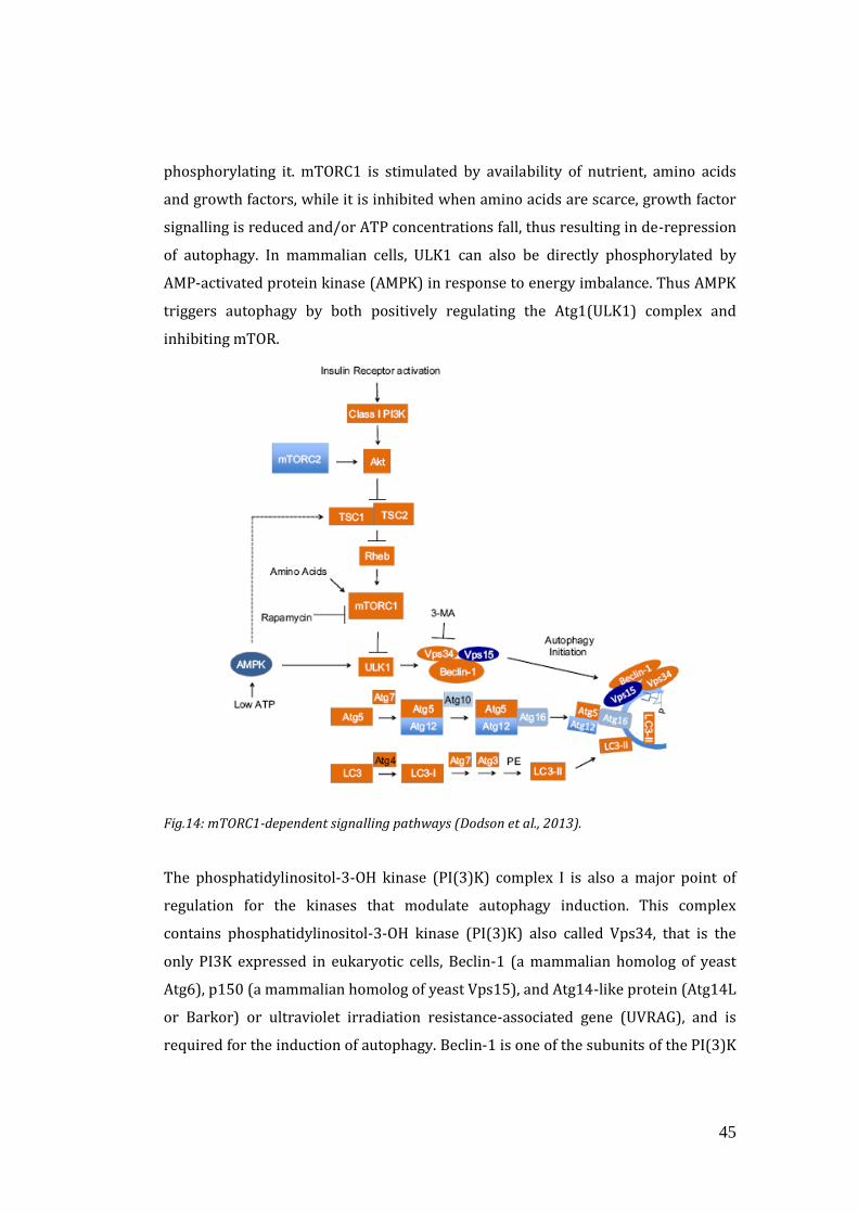
45
phosphorylating it. mTORC1 is stimulated by availability of nutrient, amino acids
and growth factors, while it is inhibited when amino acids are scarce, growth factor
signalling is reduced and/or ATP concentrations fall, thus resulting in de-repression
of autophagy. In mammalian cells, ULK1 can also be directly phosphorylated by
AMP-activated protein kinase (AMPK) in response to energy imbalance. Thus AMPK
triggers autophagy by both positively regulating the Atg1(ULK1) complex and
inhibiting mTOR.
Fig.14: mTORC1-dependent signalling pathways (Dodson et al., 2013).
The phosphatidylinositol-3-OH kinase (PI(3)K) complex I is also a major point of
regulation for the kinases that modulate autophagy induction. This complex
contains phosphatidylinositol-3-OH kinase (PI(3)K) also called Vps34, that is the
only PI3K expressed in eukaryotic cells, Beclin-1 (a mammalian homolog of yeast
Atg6), p150 (a mammalian homolog of yeast Vps15), and Atg14-like protein (Atg14L
or Barkor) or ultraviolet irradiation resistance-associated gene (UVRAG), and is
required for the induction of autophagy. Beclin-1 is one of the subunits of the PI(3)K
46
complex I and its incorporation into this complex, which is essential to stimulate
PtdIns3P synthesis, is dependent on other proteins, such as Bcl-2, 14-3-3 or the
intermediate filament protein vimentin 1 (VMP1). The phosphorylation of BECLIN1
by the death-associated protein kinase (DAPK) or phosphorylation of Bcl-2 by the c-
Jun N-terminal kinase (JNK) triggers the dissociation of the Beclin-1–Bcl-2 complex
allowing Beclin-1 to associate with the PI(3)K complex I.
Furthermore, AMPK, stimulates autophagy in response to glucose starvation by
phosphorylating BECLIN1 on a different residue that of the inhibitory kinases, and
promotes its incorporation into the PI(3)K complex I (Boya et al 2013). In this
condition, in fact, autophagy is up-regulated as compensation for the loss of key
metabolites, whereas loss of autophagy or excessive autophagy may be detrimental
to the cell (Dodson et al., 2013).
Some of the other regulatory molecules that induce autophagy are the eukaryotic
initiation factor 2α (eIF2α), which responds to nutrient starvation, double-stranded
RNA, and unfolded protein response (UPR), the major ER stress pathway
(Buchberger et al., 2010; Kouroku et al., 2007).
Furthermore, mitochondria detain an important role in autophagy regulation. I have
already reported that mitophagy is activated upon mitochondria damage. It is also
well known that damaged mitochondria produce ROS thus increasing cell oxidative
stress.
Oxidative stress reflects an imbalanced condition between reactive oxygen species
(ROS) production and the capability of the cell to buffer or eliminate these
molecules. In fact, ROS are formed by the incomplete reduction of oxygen and are
produced at low levels under normal physiological condition as a result of
mitochondria respiration and a number of other processes. The cell is able to cover
this production through several antioxidant defences. When these systems are
overloaded oxidative stress occurs and leads ultimately to cell death. Oxidative
stress is responsible for DNA mutations, and damage of cellular components such as
lipids and proteins; in particular it can lead to the non-specific post-translational
47
modifications of proteins, named carbonylation, and contributes to protein
aggregation (Underwood et al.,2010; Rossi et al., 2008; Lee et al., 2012).
Oxidative stress has been demonstrated across several tissue types to play an
important role in control of autophagy, ageing and in the progression of a multitude
of diseases including neurodegeneration, cardiovascular diseases, and cancer (Lee et
al. 2012). A general concept is that oxidative stress induces autophagy to remove
oxidized protein (Dodson et al., 2013) and dysfunctional organelles (Figure 15).
Furthermore, it has been demonstrated that anti-oxidant treatment is able to
prevent or rescue protein oxidation (Whidden et al., 2010; Desaphy et al., 2010), but
other studies pointed out its effect on autophagy. A recent work showed that both
thiol antioxidants, in particular N-acetil-cysteine (NAC), and non-thiol antioxidants,
profoundly impair both basal and induced autophagy, through different signalling
pathways (Underwood et al., 2010). In fact, although thiol antioxidants inhibit
mTOR, they also reduce the phosphorilation levels of JNK and BCL-2, reducing the
availability of BECLIN1, that is required for autophagosome formation, thus
resulting in autophagy impairment. On the contrary, non-thiol antioxidants activates
mTOR, that consequently sequesters ULK-1 from autophagy pathway (Underwood
et al., 2010). These findings suggest that basal level of oxidative stress is an
important signal that mediates autophagy activation thus maintaining tissue
homeostasis, because depletion of ‘physiological ROS’ in healthy cells leads to
impaired autophagy and homeostatic imbalance.
Oxidative stress can also induce autophagy, through p53 activation. This is a very
open topic, since there are opposite evidence regarding the role of p53, but what is
known is that p53 is a tumor suppressor protein that has a dual effect on autophagy,
acting both as a positive and negative regulator.
It was demonstrated that p53 induces autophagy via its transcriptional activity and
acts as a negative regulator of autophagy via its cytoplasmic functions (Green and
Kroemer, 2009). On the contrary, a recent work reported that cytosolic p53 leads to
autophagy induction by activation of AMPK and mTOR suppression, and inhibit
autophagy translocating to the nucleus and increasing transcription of TSC1, the
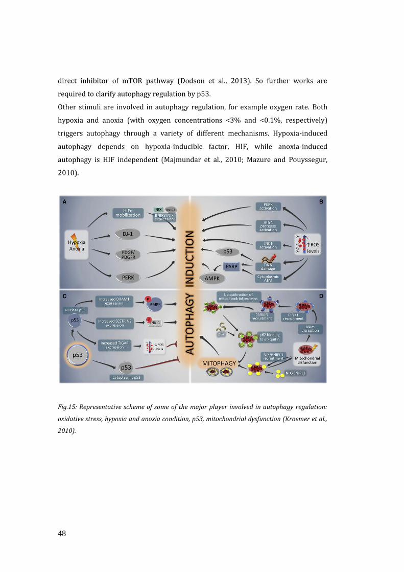
48
direct inhibitor of mTOR pathway (Dodson et al., 2013). So further works are
required to clarify autophagy regulation by p53.
Other stimuli are involved in autophagy regulation, for example oxygen rate. Both
hypoxia and anoxia (with oxygen concentrations <3% and <0.1%, respectively)
triggers autophagy through a variety of different mechanisms. Hypoxia-induced
autophagy depends on hypoxia-inducible factor, HIF, while anoxia-induced
autophagy is HIF independent (Majmundar et al., 2010; Mazure and Pouyssegur,
2010).
Fig.15: Representative scheme of some of the major player involved in autophagy regulation:
oxidative stress, hypoxia and anoxia condition, p53, mitochondrial dysfunction (Kroemer et al.,
2010).
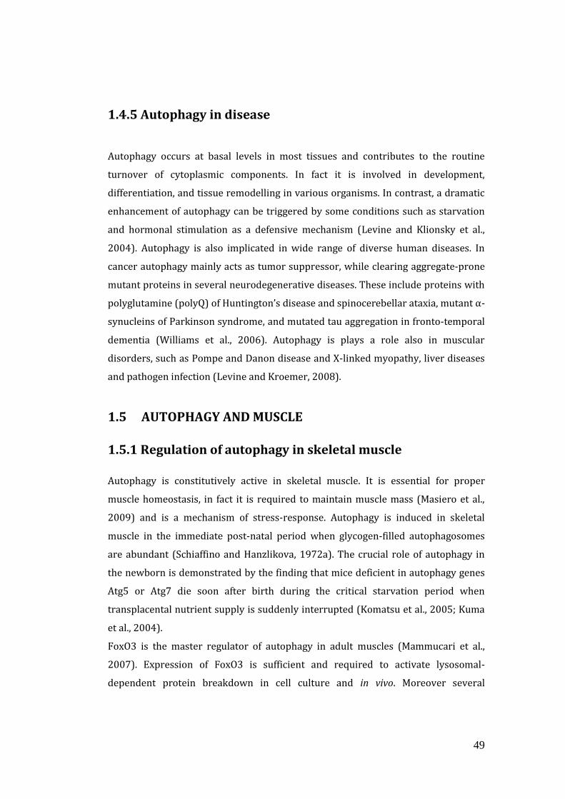
49
1.4.5 Autophagy in disease
Autophagy occurs at basal levels in most tissues and contributes to the routine
turnover of cytoplasmic components. In fact it is involved in development,
differentiation, and tissue remodelling in various organisms. In contrast, a dramatic
enhancement of autophagy can be triggered by some conditions such as starvation
and hormonal stimulation as a defensive mechanism (Levine and Klionsky et al.,
2004). Autophagy is also implicated in wide range of diverse human diseases. In
cancer autophagy mainly acts as tumor suppressor, while clearing aggregate-prone
mutant proteins in several neurodegenerative diseases. These include proteins with
polyglutamine (polyQ) of Huntington’s disease and spinocerebellar ataxia, mutant α-
synucleins of Parkinson syndrome, and mutated tau aggregation in fronto-temporal
dementia (Williams et al., 2006). Autophagy is plays a role also in muscular
disorders, such as Pompe and Danon disease and X-linked myopathy, liver diseases
and pathogen infection (Levine and Kroemer, 2008).
1.5 AUTOPHAGY AND MUSCLE
1.5.1 Regulation of autophagy in skeletal muscle
Autophagy is constitutively active in skeletal muscle. It is essential for proper
muscle homeostasis, in fact it is required to maintain muscle mass (Masiero et al.,
2009) and is a mechanism of stress-response. Autophagy is induced in skeletal
muscle in the immediate post-natal period when glycogen-filled autophagosomes
are abundant (Schiaffino and Hanzlikova, 1972a). The crucial role of autophagy in
the newborn is demonstrated by the finding that mice deficient in autophagy genes
Atg5 or Atg7 die soon after birth during the critical starvation period when
transplacental nutrient supply is suddenly interrupted (Komatsu et al., 2005; Kuma
et al., 2004).
FoxO3 is the master regulator of autophagy in adult muscles (Mammucari et al.,
2007). Expression of FoxO3 is sufficient and required to activate lysosomal-
dependent protein breakdown in cell culture and in vivo. Moreover several
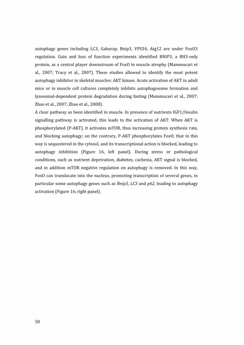
50
autophagy genes including LC3, Gabarap, Bnip3, VPS34, Atg12 are under FoxO3
regulation. Gain and loss of function experiments identified BNIP3, a BH3-only
protein, as a central player downstream of FoxO in muscle atrophy (Mammucari et
al., 2007; Tracy et al., 2007). These studies allowed to identify the most potent
autophagy inhibitor in skeletal muscles: AKT kinase. Acute activation of AKT in adult
mice or in muscle cell cultures completely inhibits autophagosome formation and
lysosomal-dependent protein degradation during fasting (Mammucari et al., 2007;
Zhao et al., 2007; Zhao et al., 2008).
A clear pathway as been identified in muscle. In presence of nutrients IGF1/Insulin
signalling pathway is activated, this leads to the activation of AKT. When AKT is
phosphorylated (P-AKT), it activates mTOR, thus increasing protein synthesis rate,
and blocking autophagy; on the contrary, P-AKT phosphorylates FoxO, that in this
way is sequestered in the cytosol, and its transcriptional action is blocked, leading to
autophagy inhibition (Figure 16, left panel). During stress or pathological
conditions, such as nutrient deprivation, diabetes, cachexia, AKT signal is blocked,
and in addition mTOR negative regulation on autophagy is removed. In this way,
FoxO can translocate into the nucleus, promoting transcription of several genes, in
particular some autophagy genes such as Bnip3, LC3 and p62, leading to autophagy
activation (Figure 16, right panel).
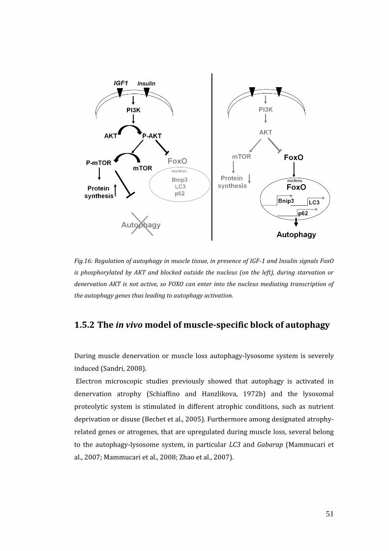
51
Fig.16: Regulation of autophagy in muscle tissue, in presence of IGF-1 and Insulin signals FoxO
is phosphorylated by AKT and blocked outside the nucleus (on the left), during starvation or
denervation AKT is not active, so FOXO can enter into the nucleus mediating transcription of
the autophagy genes thus leading to autophagy activation.
1.5.2 The in vivo model of muscle-specific block of autophagy
During muscle denervation or muscle loss autophagy-lysosome system is severely
induced (Sandri, 2008).
Electron microscopic studies previously showed that autophagy is activated in
denervation atrophy (Schiaffino and Hanzlikova, 1972b) and the lysosomal
proteolytic system is stimulated in different atrophic conditions, such as nutrient
deprivation or disuse (Bechet et al., 2005). Furthermore among designated atrophy-
related genes or atrogenes, that are upregulated during muscle loss, several belong
to the autophagy-lysosome system, in particular LC3 and Gabarap (Mammucari et
al., 2007; Mammucari et al., 2008; Zhao et al., 2007).
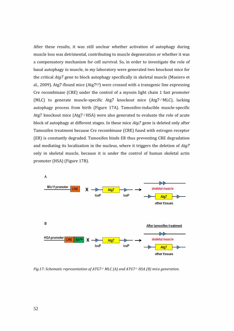
52
After these results, it was still unclear whether activation of autophagy during
muscle loss was detrimental, contributing to muscle degeneration or whether it was
a compensatory mechanism for cell survival. So, in order to investigate the role of
basal autophagy in muscle, in my laboratory were generated two knockout mice for
the critical Atg7 gene to block autophagy specifically in skeletal muscle (Masiero et
al., 2009). Atg7-floxed mice (Atg7f/f) were crossed with a transgenic line expressing
Cre recombinase (CRE) under the control of a myosin light chain 1 fast promoter
(MLC) to generate muscle-specific Atg7 knockout mice (Atg7-/-MLC), lacking
autophagy process from birth (Figure 17A). Tamoxifen-inducible muscle-specific
Atg7 knockout mice (Atg7-/-HSA) were also generated to evaluate the role of acute
block of autophagy at different stages. In these mice Atg7 gene is deleted only after
Tamoxifen treatment because Cre recombinase (CRE) fused with estrogen receptor
(ER) is constantly degraded. Tamoxifen binds ER thus preventing CRE degradation
and mediating its localization in the nucleus, where it triggers the deletion of Atg7
only in skeletal muscle, because it is under the control of human skeletal actin
promoter (HSA) (Figure 17B).
Fig.17: Schematic representation of ATG7-/- MLC (A) and ATG7-/- HSA (B) mice generation.
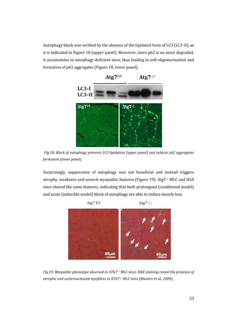
53
Autophagy block was verified by the absence of the lipidated form of LC3 (LC3-II), as
it is indicated in Figure 18 (upper panel). Moreover, since p62 is no more degraded,
it accumulates in autophagy deficient mice, thus leading to self-oligomerization and
formation of p62 aggregates (Figure 18, lower panel).
Fig.18: Block of autophagy prevents LC3 lipidation (upper panel) and induces p62 aggregates
formation (lower panel).
Surprisingly, suppression of autophagy was not beneficial and instead triggers
atrophy, weakness and several myopathic features (Figure 19). Atg7-/- MLC and HSA
mice shared the same features, indicating that both prolongued (conditional model)
and acute (inducible model) block of autophagy are able to induce muscle loss.
Fig.19: Myopathic phenotype observed in ATG7-/- MLC mice. H&E staining reveal the presence of
atrophic and centernucleated myofibres in ATG7-/- MLC mice (Masiero et al., 2009).
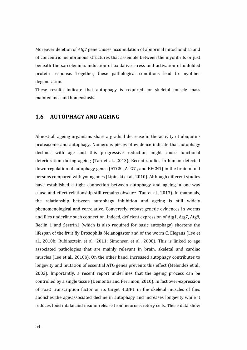
54
Moreover deletion of Atg7 gene causes accumulation of abnormal mitochondria and
of concentric membranous structures that assemble between the myofibrils or just
beneath the sarcolemma, induction of oxidative stress and activation of unfolded
protein response. Together, these pathological conditions lead to myofiber
degeneration.
These results indicate that autophagy is required for skeletal muscle mass
maintenance and homeostasis.
1.6 AUTOPHAGY AND AGEING
Almost all ageing organisms share a gradual decrease in the activity of ubiquitin-
proteasome and autophagy. Numerous pieces of evidence indicate that autophagy
declines with age and this progressive reduction might cause functional
deterioration during ageing (Tan et al., 2013). Recent studies in human detected
down-regulation of autophagy genes (ATG5 , ATG7 , and BECN1) in the brain of old
persons compared with young ones (Lipinski et al., 2010). Although different studies
have established a tight connection between autophagy and ageing, a one-way
cause-and-effect relationship still remains obscure (Tan et al., 2013). In mammals,
the relationship between autophagy inhibition and ageing is still widely
phenomenological and correlative. Conversely, robust genetic evidences in worms
and flies underline such connection. Indeed, deficient expression of Atg1, Atg7, Atg8,
Beclin 1 and Sestrin1 (which is also required for basic autophagy) shortens the
lifespan of the fruit fly Drosophila Melanogaster and of the worm C. Elegans (Lee et
al., 2010b; Rubinsztein et al., 2011; Simonsen et al., 2008). This is linked to age
associated pathologies that are mainly relevant in brain, skeletal and cardiac
muscles (Lee et al., 2010b). On the other hand, increased autophagy contributes to
longevity and mutation of essential ATG genes prevents this effect (Melendez et al.,
2003). Importantly, a recent report underlines that the ageing process can be
controlled by a single tissue (Demontis and Perrimon, 2010). In fact over-expression
of FoxO transcription factor or its target 4EBP1 in the skeletal muscles of flies
abolishes the age-associated decline in autophagy and increases longevity while it
reduces food intake and insulin release from neurosecretory cells. These data show
55
that maintenance of a normal autophagy level in skeletal muscle, but not in adipose
tissue, positively affects whole-body metabolism (Demontis and Perrimon, 2010).
FoxO3 is the a main regulator of autophagy (Mammucari et al., 2007; Mammucari et
al., 2008; Zhao et al., 2007) and, interestingly, genetic variations in the human FoxO3
have been linked to longevity in multiple population studies (Kenyon, 2010).
As I reported before, sarcopenia can be defined as ageing of muscle tissue. In
mammals the role of autophagy during sarcopenia is still controversial, because
either impaired or excessive autophagy have been associated to it (Wenz et al.,
2009; Wohlgemuth et al., 2010). Among all the studies done, loss-of-function
experiments that support the involvement of autophagy in ageing sarcopenia are
still lacking.
1.7 AUTOPHAGY AND EXERCISE
As I reported before, physical exercise is an important lifestyle practice that induces
a multitude of beneficial adaptations both in humans and rodents. Regular physical
activity has been demonstrated to improve glucose and lipid homeostasis, maintain
muscle mass and delay ageing (Melov et al., 2007; Fontana et al., 2010; Sandri et al.,
2013; Schiaffino et al., 2013; Coen et al., 2013; Toledo et al., 2013). Although the
positive effects of exercise are undisputed, the underlying mechanisms are still
under vigorous investigation, in particular the possible link with autophagy.
Previous work revealed that an acute bout of exercise is sufficient to induce
autophagy in skeletal muscle (Grumati et al., 2011). Others have further confirmed
these findings reporting that physical activity-induced adaptations that may be
mediated by the activation of autophagy (He et al., 2012; Kim et al., 2013).
Mice with defective stress-induced autophagy, but proper basal autophagy, were
found to run significantly less on a treadmill than the wild types (He et al., 2012).
Moreover, these mice do not obtain the same exercise-mediated benefits and were
not protected from high fat diet-induced glucose intolerance. This supports that
exercise-induced metabolic rejuvenation occurs through the stimulation of
autophagy. Moreover, He et al. identified AMPK as a potential player in the exercise-
induced autophagy-mediated metabolic improvements. In their hands, AMPK
56
activation presumably leads to the up-regulation of the glucose transporter, GLUT4,
at the muscle membrane, thus increasing the capacity for muscular glucose uptake
(He et al., 2012). However, the signalling cascade responsible has not been
elucidated yet. These findings remain controversial as skeletal muscle–specific
autophagy-knockout mice show the opposite phenotype. These mice appear to have
an improved metabolic profile and increased sensitivity to insulin, that protects
them from diet-induced obesity (Kim et al., 2013). These contrasting results may be
due to the difference in tissue specific versus general autophagy disturbance and,
therefore, highlight a potential cell autonomous regulation, which has yet to be
investigated.
In conclusion, it is still unknown whether it is whole body or muscle-specific
autophagy that is required to sustain contraction, maintain glucose homeostasis,
and trigger exercise-induced benefits.
57
1.8 AIM OF THE WORK
It is widely accepted that autophagy declines with age and this progressive
reduction might have a role in the functional degeneration of tissues, but a direct
causative relationship still remains undefined. Moreover, the role of autophagy
during ageing of muscle tissue, in mammals, is still controversial and loss-of-
function experiments that support its involvement are still lacking.
In order to investigate the role of autophagy inhibition in skeletal muscle during
ageing, we mimic that condition in vivo, through muscle-specific block of autophagy
process.
My work is focused on the characterization of muscle-specific aged autophagy
knockout mice (Atg7-/-MLC).
Moreover, since it is known that exercise has a beneficial effect on ageing features
and moreover it is able to reactivate autophagy in elderly, we wanted to understand
whether these beneficial effects are triggered by autophagy, and so indentify the
role of autophagy during muscle contraction. It is known that autophagy is activated
upon exercise, but the effects of this induction are still controversial. In fact, whether
it is whole body or muscle specific autophagy that is required to sustain contraction,
maintain glucose homeostasis, and triggers exercise-induced benefits, remains
unknown.
In order to clarify this issue, we used muscle-specific inducible autophagy deficient
mice (Atg7-/-HSA), to minimize the chance of any adaptations and compensations
that occur with constitutive deletion of genes.

58
59
2. MATERIALS AND METHODS
2.1 GENERATION OF MUSCLE-SPECIFIC ATG7 KNOCKOUT
MICE
Muscle-specific Atg7-/- MLC and Atg7-/- HSA mice generation have been previously
described by Masiero et al., 2009. Briefly Atg7 floxed mice Atg7f/f (Komatsu et al.,
2005) were crossed with transgenic mice expressing Cre-recombinase (CRE) under
the control of a Myosin Light Chain 1 fast promoter (MLC-1f), that is expressed only
in skeletal muscle during the embryonic development. In order to obtain inducible
muscle specific Atg7 knockout mice, Atg7 floxed mice (Komatsu et al., 2005) (Atg7f/f)
were crossed with transgenic mice expressing a Cre-recombinase fused with a
modified estrogen receptor domain (Cre-ERTM) driven by Human Skeletal Actin
promoter (HSA) (Schuler et al., 2005). In order to induce the deletion of Atg7 gene
we treated animals with Tamoxifen food (Harlan) for 9 weeks. We characterized
muscle-specific autophagy deficient (Atg7-/-) and control (Atg7f/f) mice. We analized
adult (2, 5 and 10 months old) and aged (26 months old) mice.
2.1.1 Genotyping of muscle specific Atg7 knockout mice
Mice were identified by analyzing the presence of Cre-recombinase on genomic DNA
by PCR .
We used a lysis buffer containing Tris-HCL 1M pH 7.5 and Proteinase K 10mg/mL
(Life Technologies).
The samples were denaturated by incubation for 1 hour at 57°C and then the
proteinase K was inactivated at 99°C for 5 minutes. For the PCR reaction we used
the following primers and program:
Primers:
NSP-780: CACCAGCCAGCTATCAACTCG
NSP-979: TTACATTGGTCCAGCCACCAG
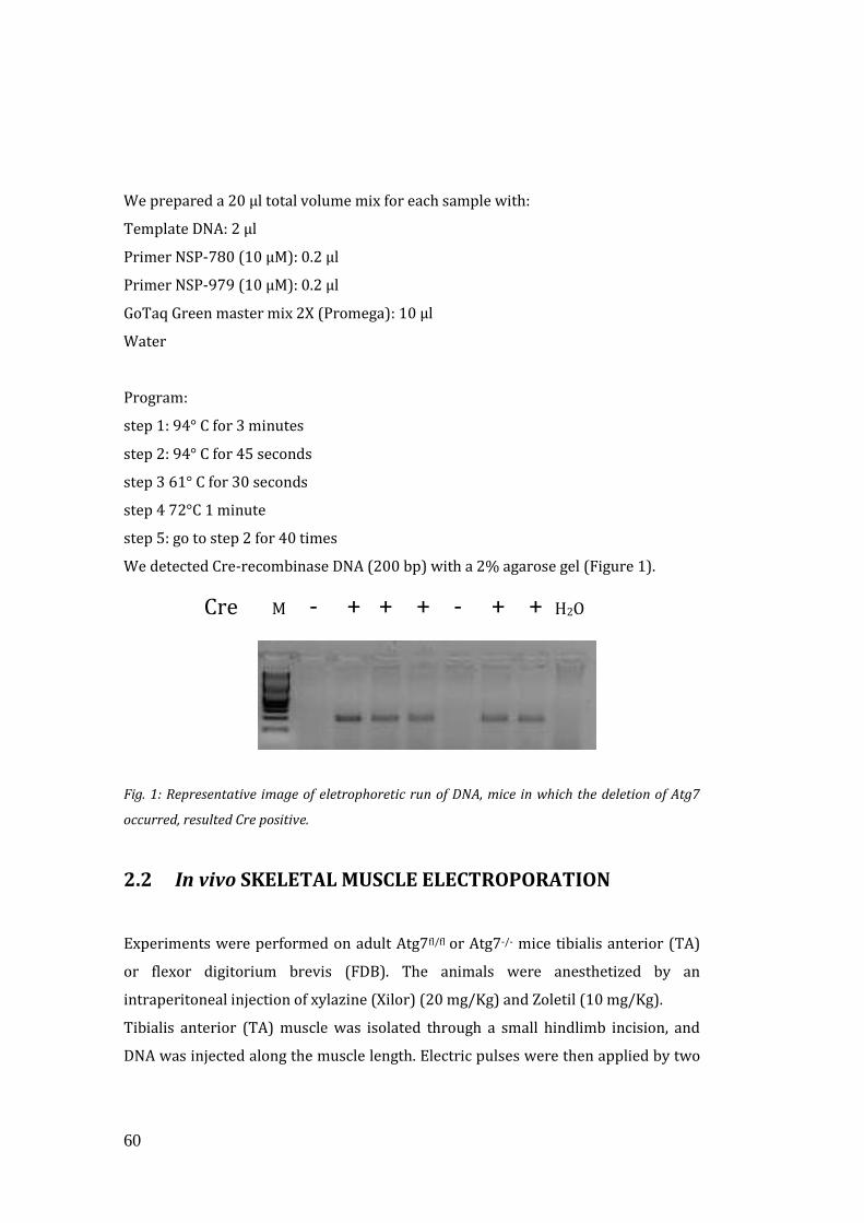
60
We prepared a 20 μl total volume mix for each sample with:
Template DNA: 2 μl
Primer NSP-780 (10 μM): 0.2 μl
Primer NSP-979 (10 μM): 0.2 μl
GoTaq Green master mix 2X (Promega): 10 μl
Water
Program:
step 1: 94° C for 3 minutes
step 2: 94° C for 45 seconds
step 3 61° C for 30 seconds
step 4 72°C 1 minute
step 5: go to step 2 for 40 times
We detected Cre-recombinase DNA (200 bp) with a 2% agarose gel (Figure 1).
Fig. 1: Representative image of eletrophoretic run of DNA, mice in which the deletion of Atg7
occurred, resulted Cre positive.
2.2 In vivo SKELETAL MUSCLE ELECTROPORATION
Experiments were performed on adult Atg7fl/fl or Atg7-/- mice tibialis anterior (TA)
or flexor digitorium brevis (FDB). The animals were anesthetized by an
intraperitoneal injection of xylazine (Xilor) (20 mg/Kg) and Zoletil (10 mg/Kg).
Tibialis anterior (TA) muscle was isolated through a small hindlimb incision, and
DNA was injected along the muscle length. Electric pulses were then applied by two
Cre M - + + + - + + H2O
61
stainless steel spatula electrodes placed on each side of the isolated muscle belly (50
Volts/cm, 5 pulses, 200 ms intervals). Muscles were analyzed 14 days later. No
evidence of necrosis or inflammation as a result of the transfection procedure were
observed (Sandri et al., 2004 and Donà et al., 2003).
Tibialis Anterior of Atg7-/- MLC mice were transfected with FGFBP1-V5 (20 μg) and
GFP (5 μg) plasmids. Tibialis Anterior of control mice were transfected with oligos
(25 μg) for knocking down experiments.
FDB electroporation did not require muscle isolation. We injected 5 μl of
Hyaluronidase (2mg/ml)(Sigma-Aldrich) in the feet of anesthetized mice, to soften
muscle tissue underneath the epidermis. We waited 50 minutes to inject DNA and
after 10 minutes electric pulses were applied by two stainless needles placed at one
cm from each other (100V/cm) (100 Volts/cm, 20 pulses, 1 s intervals). Muscles
were analyzed 7 days later. No evidence of necrosis or inflammation were observed
after the transfection procedure. FDB of Atg7f/f HSA and Atg7-/- HSA mice were
transfected with roGFP plasmid (15 μg).
2.3 MEASUREMENTS OF MUSCLE FORCE IN VIVO
Muscle force was measured in a living animal as previously described (Blaauw et al.,
2008). We performed this experiment in collaboration with Bert Blaauw (VIMM,
Padua). Briefly gastrocnemius (GCN) muscle contractile performance was measured
in vivo using a 305B muscle lever system (Aurora Scientific Inc.) in anaesthetized
mice. Contraction was elicited by electrical stimulation of the sciatic nerve. The
torque developed during isometric contractions was measured at stepwise
increasing stimulation frequency, with pauses of at least 30 seconds between stimuli
to avoid effects due to fatigue. Duration of the trains never exceeded 600 ms. Force
developed by plantar flexor muscles was calculated by dividing torque by the lever
arm length (taken as 2.1 mm).
62
2.4 HISTOLOGY ANALYSES AND FIBRE SIZE
MEASUREMENTS
Muscles were collected and directly frozen by immersion in liquid nitrogen. Then we
cut muscle cryosections by using Cryostat (Leica CM 1950), 10μm thick for histology
and 7μm thick for immunostaining analyses. TA Cryosections, 10μm thick, were
used to analyze tissue morphology with different methods, listed below. Images
were collected with an epifluorescence Leica DM5000B microscope, equipped with a
Leica DFC300-FX digital charge-coupled device camera, by using Leica DC Viewer
software.
For electron microscopy, we used conventional fixation-embedding procedures
based on glutaraldehyde-osmium fixation and Epon embedding.
2.4.1 Haematoxylin and Eosin staining (H&E)
Haematoxylin colors basophilic structures that are usually the ones containing
nucleic acids, such as ribosomes, chromatin-rich cell nuclei, and the cytoplasmc
regions rich in RNA. Eosin colors eosinophilic structures bright pink. The
eosinophilic structures are generally intracellular or extracellular protein.
The methods consist of:
Materials Time
3 washes in PBS 5 minutes each
Harris Haematoxylin (Sigma-Aldrich) 1 minute
Wash in running tap water 3 minutes
Eosin Y Solution Alcoholic (Sigma-Aldrich) 1 minute
Ethanol 50% 30 seconds
Ethanol 70% 30 seconds
63
Ethanol 100% 5 minutes
Ethanol 100% 10 minutes
Xilen 3 minutes
Mount with Eukitt (Sigma-Aldrich)
2.4.2 Succinate dehydrogenase (SDH)
The succinate dehydrogenase is an enzyme complex, bound to the inner
mitochondrial membrane. With this staining it is possible to evaluate approximately
the quantity of mitochondria present in the muscle fibres, through colorimetric
evaluation. The reaction gives a purple coloration in the oxidative fibres. The
sections were incubated for 30 minutes at 37°C with SDH solution (0.2M sodium
succinate) (Sigma-Aldrich), 0.2M phosphate buffer (Sigma) ph 7.4 and 50mg of nitro
blue tetrazolium (NBT)(Sigma-Aldrich). After the incubation, the sections were
washed 3 minutes with PBS and then mounted with Mounting medium (Dako).
2.4.3 Fibre Cross-Sectional Area (CSA)
Fibre Cross-Sectional Area was measured, by using ImageJ software (National
Institutes of Health). All data are expressed as the mean SEM (error bars).
Comparisons were made by using t test, with *P<0.05 being considered statistically
significant.
2.5 IMMUNOHISTOCHEMISTRY ANALYSES
TA muscle cryosections, 7 µm thick, were processed for immunostaining. All data
are expressed as the mean SEM (error bars). Comparisons were made by using t
test, with *P<0.05 being considered statistically significant.
64
2.5.1 NCAM staining
Muscle cryosections were fixed with Methanol -20°C, treated with 0,1% Triton for 5
minutes and incubated in blocking solution (0.5% BSA, 10% goat serum in PBS) at
room temperature (RT) for 40 minutes. Samples were then incubated with the
primary antibody against NCAM (Millipore) (dilution 1:200) at 4°C over-night. Then
the sections were washed with PBS three times for 5 minutes and incubated with
the anti-rabbit-Cy3-conjugated secondary antibodies (dilution 1:200) at 37°C for 1
hour (Life Technologies). After the washes and incubation with DAPI, that labels
nuclei, slides were mounted with mounting medium (Dako).
2.5.2 MuSK staining
Cryosections were fixed with PFA 4%, treated with 0,1% Triton for 5 minutes and
incubated in blocking solution (0.5% BSA, 10% mouse serum in PBS) at RT for 40
minutes. After that samples were incubated with the primary antibody against MuSK
(kind gift from M. Ruegg), at 4°C over-night (dilution 1:200). Sections were then
washed with PBS three times for 5 minutes and incubated with the anti-rabbit-Cy3-
conjugated secondary antibodies (dilution 1:200) at 37°C for 1hour (Life
Technologies). After the washes, sections were incubated with anti-Bungarotoxin-
Alexa 488 (dilution 1:500) from Life Technologies at 37°C for 1 hour. After the
washes and incubation with DAPI, slides were mounted with mounting medium
(Dako).
2.5.3 IgG staining
Cryosections were incubated in blocking solution (0.5% BSA, 10% goat serum in
PBS) at RT for 20 minutes. Samples were then incubated with anti-mouse-Cy3-
conjugated secondary antibodies (dilution 1:200) at 37°C for 1hour (Life
Technologies). After the wash and incubation with DAPI, slides were mounted with
mounting medium (Dako).
65
2.6 IMMUNOBLOTTING
Cryosections of 20 μm of TA muscles were lysed with 100 μl of a buffer containing
50 mM Tris pH 7.5, 150 mM NaCl, 10 mM MgCl2, 0.5 mM DTT, 1 mM EDTA, 10%
glycerol, 2% SDS, 1% Triton X-100, Roche Complete Protease Inhibitor Cocktail and
Sigma Protease Inhibitor Cocktail. After incubation at 70°C for 10 minutes and
centrifugation at 11000 g for 10 minutes at 4°C, the surnatant protein concentration
was measured using BCA protein assay kit (Pierce) following the manufacturer’s
instructions.
2.6.1 Protein gel Electrophoresis
The extracted proteins from TA muscle were solubilized in Loading buffer composed
by 5μl of 4X NuPAGE® LDS Sample Buffer (Life Technologies) and 1μl of 20X DTT
(Life Technologies). The volume of each sample was brought to 20μl with TBS 1X.
The samples were denaturated at 70°C for 10 minutes.
Samples were loaded on SDS 4-12% precast polyacrylamide gels (NuPAGE Novex-
Bis-tris-gels) or in SDS 3-8% depending on the protein to be analyzed (Life
Technologies). The electrophoresis was run in 1X MES Running buffer or 1X Tris-
Acetate Running buffer respectively (Life Technologies) for 1 hour and 30 minutes
at 150V constant.
2.6.2 Transfer of the protein to the PVDF membrane
After the electrophoretic run, proteins were transferred from gels to PVDF
membranes, previously activated with methanol. The gel and the membrane were
equilibrated in Transfer Buffer. The Transfer Buffer was prepared as follows: 50 ml
of 20X NuPAGE® Transfer buffer (Life Technologies), 1 ml of 10X NuPAGE®
Antioxidant (Life Technologies), 200 ml of 20% Methanol (Sigma-Aldrich). The
volume was brought to 1l with distilled water. The blotting was obtained by
applying a current of 400mA for two hours at 4°C. To evaluate the efficiency of the
transfer, proteins were stained with Red Ponceau 1x (Sigma-Aldrich). The staining
was easily reversed by washing with distilled water.
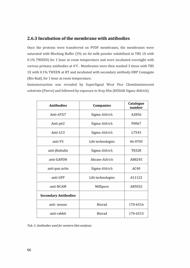
66
2.6.3 Incubation of the membrane with antibodies Once the proteins were transferred on PVDF membranes, the membranes were
saturated with Blocking Buffer (5% no fat milk powder solubilized in TBS 1X with
0.1% TWEEN) for 1 hour at room temperature and were incubated overnight with
various primary antibodies at 4°C . Membranes were then washed 3 times with TBS
1X with 0.1% TWEEN at RT and incubated with secondary antibody-HRP Conjugate
(Bio-Rad), for 1 hour at room temperature.
Immunoreaction was revealed by SuperSignal West Pico Chemiluminescent
substrate (Pierce) and followed by exposure to Xray film (KODAK Sigma-Aldrich).
Antibodies Companies Catalogue number
Anti-ATG7 Sigma-Aldrich A2856
Anti-p62 Sigma-Aldrich P0067
Anti-LC3 Sigma-Aldrich L7543
anti-V5 Life technologies 46-0705
anti-βtubulin Sigma-Aldrich T8328
anti-GAPDH Abcam-Aldrich AB8245
anti-pan actin Sigma-Aldrich AC40
anti-GFP Life technologies A11122
anti-NCAM Millipore AB5032
Secondary Antibodies
anti- mouse Biorad 170-6516
anti-rabbit Biorad 170-6515
Tab. 1: Antibodies used for western blot analyses.
67
All the peroxidase-conjugated secondary antibodies were from Bio-Rad. Blots were
stripped using Stripping Solution, containing 25mM glycine and 1% SDS, pH 2.
2.7 FUNCTIONAL ASSAYS ON SINGLE MUSCLE FIBRES
All these experiments were done in collaboration with the group of Prof. Bottinelli
(Physiology department, University of Pavia).
2.7.1 Single fibre dissection and experimental set-up
The method used for single muscle fibres dissection, the solutions and the
experimental set-up have been previously described (Bottinelli et al., 1996). Briefly,
after animal sacrifice, muscle bundles were prepared and stored at -20°C. On the day
of the experiment, single muscle fibres were dissected from a bundle with the help
of a stereomicroscope. Segments of muscle fibres were chemically skinned and
attached in the experimental setup. Relaxing (5mM EGTA pCa 9.0), pre-activating
(0.5mM EGTA pCa 9.0) and activating (pCa 4.5) solutions were prepared as
previously described (Bottinelli et al., 1996). The set up was placed on the stage of
an inverted microscope (Axiovert 10, Zeiss, Germany) which allowed us to view the
fibre at x 320 magnification. In this way, we measured the force generated by each
fiber and their speed of contraction. The signals from the force and displacement
transducer were fed into a personal computer and analyzed by a data analysis
software (Spike 2, CED,Cambridge, UK).
2.7.2 Single fibre analysis
Single fibre analysis was performed as previously described (Bottinelli et al., 1996).
The experiments were performed at the temperature of 12°C. Sarcomere length was
determined and set at 2.5µm. The cross sectional area (CSA) of the fibres was
determined without correction for swelling. Absolute (Po) and specific force
(Po/CSA) of the fibres were determined. At the end of the mechanical experiment
fibres were put in 20 μl of Laemmli buffer and stored at –20 °C for subsequent
electrophoretic analysis of myosin heavy chains (MHC) isoform content.
68
2.7.3 Contractile proteins for IVMA
Myosin extraction from bulk muscles
Myosin isoform 2B was extracted from bulk gastrocnemius muscles of mice
according a procedure previously described in detail (Canepari et al., 2000) and
used to prepare heavy meromyosin fraction (HMM).
Heavy meromyosin (HMM) preparation
HMM was obtained by a proteolytic digestion with -chimotrypsin of myosin
according to a modification of the method of Margossian and Lowey (Margossian
and Lowey, 1982) previously described in detail (Canepari et al., 2000).
Actin preparation
G Actin was extracted as described by Pardee and Spudich (Pardee and Spudich,
1982) from acetone powder prepared from the residues of mice muscles after
myosin extraction. After polymerization, F actin was labeled by incubation for
several hours with rhodamine-phalloidine (Molecular Probes R415) as described by
Kron et al (Kron et al., 1991).
2.7.4 In Vitro Motility Assay (IVMA)
In this assay actin and myosin are purified, then actin is labeled with rhodamine
falloidine and allowed to move over myosin molecules that were previously linked
onto a glass slide. When ATP is added, the actin-myosin complexes move out from
the in rigor state and the actin movement and velocity of sliding on myosin is
measured.
Myosin (or HMM ) was diluted to 0.1 mg/ml in a high (or low) ionic strength buffer
and infused in a flow cell treated with nitrocellulose and prepared according to
Anson et al (Anson et al., 1995). The IVMA analysis was performed according to
Canepari et al. (Canepari et al., 2000; Canepari et al., 1999) at the temperature of
25°C. The composition of the experimental buffer was MOPS 25mM (pH=7.4 at
25°C), KCl 25mM, MgCl2 4mM, EGTA 1mM, DTT 1mM, glucose oxidase 200 μ/ml,
catalase 36μg/ml, glucose 5 mg/ml, ATP 2mM. Average velocities of actin filaments
69
were determined for each myosin and HMM sample, the velocities of at least 50
filaments were measured and their distribution characterized according to
parametric statistics.
2.8 IN VIVO MICROSCOPY AND ANALYSIS OF ACHR
TURNOVER AND NMJ FRAGMENTATION
We performed these experiments in collaboration with the group of Dr. Rudolf (KIT-
Karlsruhe Institute of Technology, Karlsruhe). Bungarotoxin-Alexa 488 (1:100
dilution from 1mg/ml stock solution) (Life Technologies) was injected in the TA of
an anesthetized mice, then after 10 days, a second injection of Bungarotoxin-Alexa
594 was performed in the same TA muscle of each mice. Mice that were previously
transfected with specific plasmids had the first injection after 5 days of transfection.
In vivo microscopy of mice was performed under anesthesia using Xylazine (Xilor)
(20 mg/Kg) and Zoletil (10 mg/Kg) on a Leica SP2 confocal microscope equipped
with a 63x 1.2 N.A. water immersion objective, essentially as described previously
(Roder et al., 2010; Roder et al., 2008). Automated analysis of AChR turnover and
NMJ fragmentation used algorithms described earlier (Roder et al., 2010).
2.9 GENE EXPRESSION ANALYSIS
Quantitative Real-time PCR was performed with SYBR Green chemistry (Applied
Biosystems). SYBR green is a fluorescent dye that intercalates into double-stranded
DNA and produces a fluorescent signal. The Real-Time PCR Instrument allows real
time detection of PCR products as they accumulate during PCR cycles and create an
amplification plot, which is the plot of fluorescence signal versus cycle number. In
the initial cycles of PCR, there is little change in fluorescence signal. This defines the
baseline for the amplification plot. An increase in fluorescence above the baseline
indicates the detection of accumulated PCR products. A fixed fluorescence threshold
can be set above the baseline. The parameter Ct (threshold cycle) is defined as the
fractional cycle number at which the fluorescence passes the fixed threshold. So the
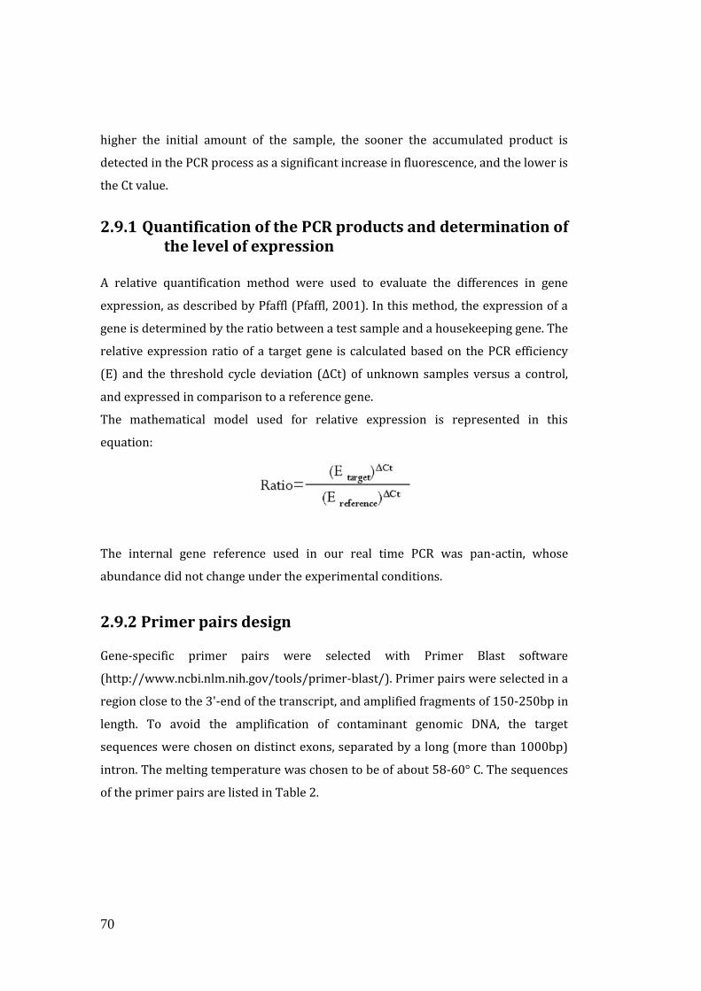
70
higher the initial amount of the sample, the sooner the accumulated product is
detected in the PCR process as a significant increase in fluorescence, and the lower is
the Ct value.
2.9.1 Quantification of the PCR products and determination of the level of expression
A relative quantification method were used to evaluate the differences in gene
expression, as described by Pfaffl (Pfaffl, 2001). In this method, the expression of a
gene is determined by the ratio between a test sample and a housekeeping gene. The
relative expression ratio of a target gene is calculated based on the PCR efficiency
(E) and the threshold cycle deviation (∆Ct) of unknown samples versus a control,
and expressed in comparison to a reference gene.
The mathematical model used for relative expression is represented in this
equation:
The internal gene reference used in our real time PCR was pan-actin, whose
abundance did not change under the experimental conditions.
2.9.2 Primer pairs design Gene-specific primer pairs were selected with Primer Blast software
(http://www.ncbi.nlm.nih.gov/tools/primer-blast/). Primer pairs were selected in a
region close to the 3'-end of the transcript, and amplified fragments of 150-250bp in
length. To avoid the amplification of contaminant genomic DNA, the target
sequences were chosen on distinct exons, separated by a long (more than 1000bp)
intron. The melting temperature was chosen to be of about 58-60° C. The sequences
of the primer pairs are listed in Table 2.
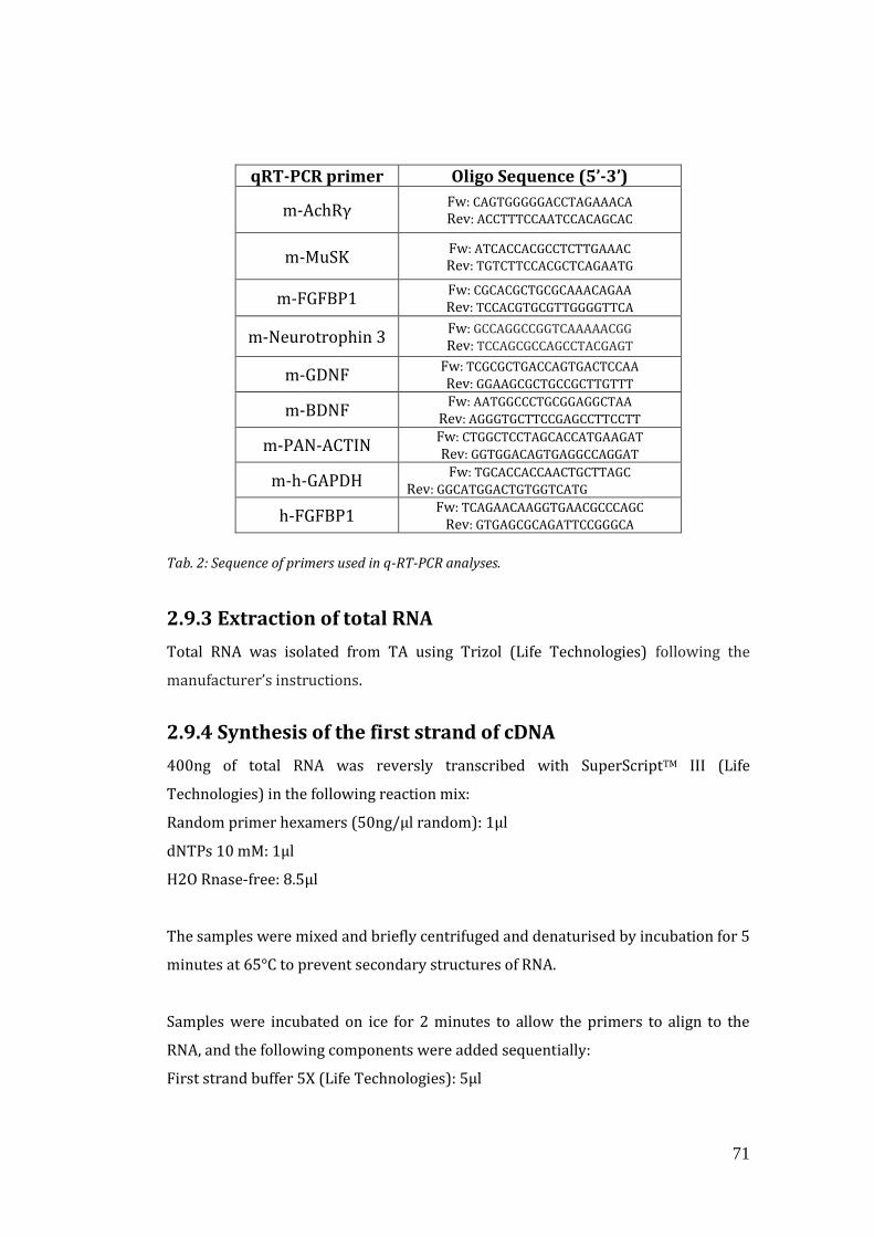
71
qRT-PCR primer Oligo Sequence (5’-3’)
m-AchRγ Fw: CAGTGGGGGACCTAGAAACA
Rev: ACCTTTCCAATCCACAGCAC
m-MuSK Fw: ATCACCACGCCTCTTGAAAC
Rev: TGTCTTCCACGCTCAGAATG
m-FGFBP1 Fw: CGCACGCTGCGCAAACAGAA
Rev: TCCACGTGCGTTGGGGTTCA
m-Neurotrophin 3 Fw: GCCAGGCCGGTCAAAAACGG
Rev: TCCAGCGCCAGCCTACGAGT
m-GDNF Fw: TCGCGCTGACCAGTGACTCCAA
Rev: GGAAGCGCTGCCGCTTGTTT
m-BDNF Fw: AATGGCCCTGCGGAGGCTAA
Rev: AGGGTGCTTCCGAGCCTTCCTT
m-PAN-ACTIN Fw: CTGGCTCCTAGCACCATGAAGAT
Rev: GGTGGACAGTGAGGCCAGGAT
m-h-GAPDH Fw: TGCACCACCAACTGCTTAGC
Rev: GGCATGGACTGTGGTCATG
h-FGFBP1 Fw: TCAGAACAAGGTGAACGCCCAGC
Rev: GTGAGCGCAGATTCCGGGCA
Tab. 2: Sequence of primers used in q-RT-PCR analyses.
2.9.3 Extraction of total RNA
Total RNA was isolated from TA using Trizol (Life Technologies) following the
manufacturer’s instructions.
2.9.4 Synthesis of the first strand of cDNA
400ng of total RNA was reversly transcribed with SuperScriptTM III (Life
Technologies) in the following reaction mix:
Random primer hexamers (50ng/μl random): 1μl
dNTPs 10 mM: 1μl
H2O Rnase-free: 8.5μl
The samples were mixed and briefly centrifuged and denaturised by incubation for 5
minutes at 65°C to prevent secondary structures of RNA.
Samples were incubated on ice for 2 minutes to allow the primers to align to the
RNA, and the following components were added sequentially:
First strand buffer 5X (Life Technologies): 5μl
72
DTT 100mM: 2μl
RNase Out (Life Technologies): 1μl
SuperScriptTM III (Life Technologies): 0.5μl
The volume was adjusted to 20ul with RNase free water.
The used reaction program was:
step1: 25°C for 10 minutes
step2: 42°C for 50 minutes
step3: 70°C for 15 minutes
At the end of the reaction, the volume of each sample was adjusted to 50ul with
RNase free water.
2.9.5 Real-Time PCR reaction 1μl of diluted cDNAs was amplified in 10μl PCR reactions in a ABI Prism 7000
(Applied Biosystem) thermocycler, coupled with a ABI Prism 7000 Sequence
Detection System (Applied Biosystems) in 96-wells plates (Micro Amp Optical,
Applied Biosystems). In each well 5ul Sample mix and 5ul reaction mix were mixed.
Sample mix was prepared as follows for 5 μl total volume:
Template cDNA: 1 μl
H2O Rnase-free: 4 μl
The SYBR Green qPCR (Applied Biosystem) was used for the Real-Time PCR reaction
as follows:
SYBR Green qPCR (Applied Biosystem): 4.8μl
Mix Primer forward /reverse 50mM: 0.2μl
The PCR cycle used for the Real-Time PCR was:
step 1: 95° C for 15 minutes
step2: 95° C for 25 seconds
step3: 58° C for 1 minute
step4: go to step 2 for 40 times
73
2.10 PLASMID CLONING
The cloning strategy required insertion of the DNA of intrest in a specific plasmid.
Then competent bacteria (Top 10) (Life Technologies) were transformed and the
plasmid was purified first with Mini-prep kit (Machery-Nagel) and then, to increase
the quantity of DNA, in particular for in vivo transfection, we prepared Maxi-prep
(Qiagen) following the manufacturer’s instructions. Final DNA concentration was
quantified by Nanovue Plus (GE Haelthcare).
2.10.1 FGFBP1 cloning
FGFBP1 was amplified from mouse cDNA by PCR using the following primers Fw:
ACCATGAGACTCCACAGCCTC and Rev: GCATGATGTCGCCTGTAACAT. The PCR
fragments then were cloned into pcDNA3.1-V5-HISTOPO vector (Life Technologies).
2.10.2 In Vivo RNAi
Oligos were cloned into Life Technologies BLOCK-IT Pol II miR RNAi Expression
Vectors. For validation of shRNA constructs, C2C12 cells were maintained in
DMEM/10%FBS and transfected with shRNA constructs using Lipofectamine 2000
(Life Technologies) according to manufacturer’s instructions. Cells were lysed 24
hours later, and immunoblotting was performed as described below. The sequences
of the Oligos Used for shRNA Production are listed in the Table 1:
shRNA oligos
Oligo sequence (5’-3’)
FGFBP1-
oligo 1 Fw: TGCTGTATTCTGGGCCTTCCCTAACGGTTTTGGCCACTGACTGACCGTTAGGGGGCCCAGAATA Rev: CCTGTATTCTGGGCCCCCTAACGGTCAGTCAGTGGCCAAAACCGTTAGGGAAGGCCCAGAATAC
FGFBP1-
oligo 2 Fw: TGCTGTGCACTGGACCTTCAGGCTGAGTTTTGGCCACTGACTGACTCAGCCTGGGTCCAGTGCA Rev: CCTGTGCACTGGACCCAGGCTGAGTCAGTCAGTGGCCAAAACTCAGCCTGAAGGTCCAGTGCAC
FGFBP1-
oligo 3 Fw: TGCTGTTCTGAGAACGCCTGAGTAGCGTTTTGGCCACTGACTGACGCTACTCACGTTCTCAGAA Rev: CCTGTTCTGAGAACGTGAGTAGCGTCAGTCAGTGGCCAAAACGCTACTCAGGCGTTCTCAGAAC
FGFBP1- oligo 4
Fw:TGCTGTTAGCATGATGTCGCCTGTAAGTTTTGGCCACTGACTGACTTACAGGCCATCATGCTAA Rev: CCTGTTAGCATGATGGCCTGTAAGTCAGTCAGTGGCCAAAACTTACAGGCGACATCATGCTAAC
74
MuSK- oligo 1
Fw: TGCTGAAATATGGCAGTCTTGTGCAGGTTTTGGCCACTGACTGACCTGCACAACTGCCATATTT Rev: CCTGAAATATGGCAGTTGTGCAGGTCAGTCAGTGGCCAAAACCTGCACAAGACTGCCATATTTC
MuSK- oligo 2
Fw: TGCTGTTAGGTTTCATCTTCACTTGCGTTTTGGCCACTGACTGACGCAAGTGAATGAAACCTAA Rev: CCTGTTAGGTTTCATTCACTTGCGTCAGTCAGTGGCCAAAACGCAAGTGAAGATGAAACCTAAC
Tab. 3: Sequence shRNA used for in vivo transfection
2.10.3 Cell culture and transient transfection C2C12 myogenic cell line were cultured in DMEM (GIBCO-Life Technologies)
supplemented with 10% foetal bovine serum. Myoblasts were transfected using
Lipofectamine 2000 (Life Technologies) according to the manufacturer’s
instructions.
2.11 PROTEIN CARBONYLS DETECTION
We used the Oxyblot assay to evaluate ongoing protein carbonylation in skeletal
muscle. Extensor Digitorum Longus (EDL) muscles were lysed in lysis buffer with
50mM of DTT. The composition of the lysis buffer is similar to that used for protein
extraction. The samples were then incubated at 70°C for 10 minutes in a
termomixer, and centrifuged at 11000 g for 10 minutes at 4°C. Then 15 µg of protein
lysates were derivatized with 2,4dinitrophenylhydrazine (DNPH) solution and then
neutralized. This way the samples can be separated on SDS-PAGE (12% (v/v)
polyacrylamide) and electro blotted onto nitrocellulose membranes. After blocking,
membranes were incubated with anti-DNP antibody (Sigma-Aldrich), washed, and
incubated with peroxidase conjugated anti-rabbit IgG antibodies. Immunoreaction
was revealed by SuperSignal West Pico Chemiluminescent substrate (Pierce) and
followed by exposure to Xray film (KODAK Sigma-Aldrich). Quantification analysis
was performed with ImageJ Software and all values were normalized for the
housekeeping gapdh.
75
2.12 EXERCISE PROTOCOL
Atg7f/f and Atg7-/- HSA mice performed one day of concentric exercise on a treadmill
(LE 8710 Panlab Technology 2B, Biological Instruments), with 10 degrees incline, at
increasing velocity, according to the protocol of acute exercise previously described
(He et al., 2012).
The eccentric training protocol consisted of 3 days running to exhaustion, with a 10
degree decline, at increasing velocity, according to the protocol of exercise
previously described (He et al., 2012).
Briefly, exercise consists in 17 cm/sec for 40 minutes, 18 cm/sec for 10 min, 20
cm/sec for 10 min, 22 cm/sec for 10 min, and then increasing velocity of 1cm/sec
and or 2 cm/sec alternatively every 5 minutes, until they were exhausted.
Exhaustion was defined as the point at which mice spent more than 5 s on the
electric shocker without attempting to resume running.
Total running distance and time were recorded for each mouse. All procedures are
specified in the projects approved by the Italian Ministero Salute, Ufficio VI
(authorization numbers C65).
2.13 ANTI-OXIDANT TREATMENT Atg7f/f and Atg7-/- MLC mice were intraperitoneally injected with Trolox (Sigma-
Aldrich) 30mg/kg daily for four weeks. Trolox is a general anti-oxidant because it is
an analogue of vitamin E.
Females Atg7f/f and Atg7-/- HSA mice were treated with N-Acetyl-Cysteine (NAC)
(Sigma-Aldrich A9165), for 6 weeks. We used 1% NAC drinking water for 5 weeks
and 2% NAC drinking water for the last week. The treatment was also maintained
during exercise training. NAC is a general anti-oxidant because it is a thiol
antioxidant.
Another group of females Atg7f/f and Atg7-/- HSA mice were then treated with
intraperitoneal injections of Mito-TEMPO (Enzo Life Science) 1.4 mg/kg daily for 7
days. Mito-TEMPO is a specific anti-oxidant, because it is a mitochondria-targeted
superoxidant dismutase mimetic (Dikalova et al., 2010).
76
2.14 ANALYSES OF MITOCHONDRIAL MEMBRANE
POTENTIAL IN ISOLATED SINGLE FIBRES
Mitochondrial membrane potential was measured in isolated fibres from flexor
digitorum brevis (FDB) muscles. FDB muscle was incubated in a DMEM (GIBCO-Life
Technologies) and Collagenase (4mg/ml)(GIBCO-Life Technologies) solution for
about one hour and 45 minutes at 37°C. Then single fibres were dissected with the
pipet with the help of a stereomicroscope. Mitochondrial membrane potential was
measured by epifluorescence microscopy based on the accumulation of TMRM
fluorescence as previously described (Romanello et al., 2010). Single fibres were
incubated with TMRM (Tetramethylrhodamine, methyl ester)(Life Technologies) a
fluorescent lipophilic cationic molecule that accumulates in mitochondria in a
potential-dependent manner. The solution contains also glucose (3,5 g/l), to sustain
the fibres during the experiment and Cyclosporin H (8mM) (Enzo Life Science), to
block mitochondrial pumps, that would transport TMRM outside the mitochondria.
In this experiment we need to perturbed the system with Oligomycin (4 µM)(Sigma-
Aldrich), that is an ATP-syntase inhibitor, because ATP-synthase can reversely
transport protons across the inner mitochondrial membrane, so maintaining the
potential also in dysfunctional mitochondria. For this reason, only this treatment
allow us to detect real dysfunctional mitochondria, that would inevitably dissipate
the potential, loosing TMRM signal. At the end, we add an uncoupling agent FCCP
(Trifluorocarbonylcyanide Phenylhydrazone) (4 µM)(Sigma-Aldrich), as
experimental control. All the images were analyzed with ImageJ Software, and we
considered fibres as depolarized when they have lost more than 10% of the initial
value of TMRM fluorescence.
2.15 MITOCHONDRIAL OXIDATIVE STRESS MEASUREMENT
Mt-roGFP1 is an indicator of mitochondrial redox status (Dooley et al., 2004; Hanson
et al., 2004). It measures the thiol/disulfide equilibrium in the mitochondrial matrix.
We transfected FDB muscles of females Atg7f/f and Atg7-/- HSA mice with mt-roGFP
77
plasmid, that is targeted to the mitochondria, one week before exercise. Then we
measured the fluorescence in single muscle fibres, that were isolated prior to and
after exercise. Mt-roGFP1 fluorescence (excitation: 405 and 480 nm, emission: 535
nm, 20X objective) was measured for 5 minutes every 10 seconds. The ratio of
fluorescence intensities (exc. 405/480) were computed. Records were analyzed
with ImageJ Software.
2.16 BLOOD METABOLITES QUANTIFICATION
This analysis was performed in collaboration with the group of Prof. Avogaro
(Venetian Institute of molecular medicine, VIMM, Padova). Blood samples were
collected before exercise and immediately after the 3 days of exercise. Blood was
collected from the orbital sinus in heparin-coated pasteur pipettes and centrifuged
immediately after collection. Plasma samples were kept at -20°C until dosing.
Blood glucose and lactate levels were measured with an YSI 2300 STAT Plus™
Glucose & Lactate Analyzer (YSI Life Sciences, Yellow Springs, OH) according to the
manufacture's instruction. Free fatty acids (FFAs) and beta-hydroxybutyrate were
dosed using an automated spectrophotometer Cobas Fara II (Roche) according to
the manufacture’s instruction.
2.17 STATISTICAL ANALYSES
Survival probability evaluated by using Kaplan-Meier method. Comparison of Atg7f/f
and Atg7-/- survival curves was performed by both Mantel-Cox and Gehan-Breslow-
Wilcoxon tests.
Comparisons were made by using t test, with p<0.05 being considered statistically
significant (*p<0.05, **p<0.01, ***p<0.001). Values are indicated in the graphs by
mean +/- standard error.

78
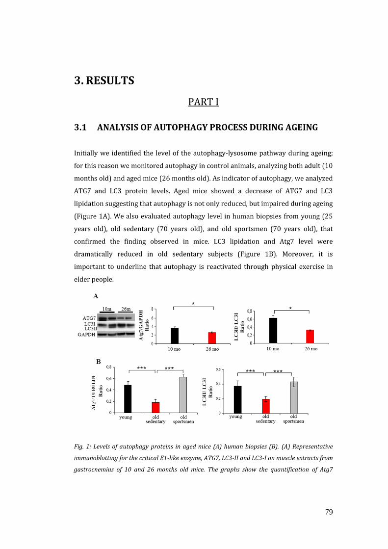
79
3. RESULTS
PART I
3.1 ANALYSIS OF AUTOPHAGY PROCESS DURING AGEING
Initially we identified the level of the autophagy-lysosome pathway during ageing;
for this reason we monitored autophagy in control animals, analyzing both adult (10
months old) and aged mice (26 months old). As indicator of autophagy, we analyzed
ATG7 and LC3 protein levels. Aged mice showed a decrease of ATG7 and LC3
lipidation suggesting that autophagy is not only reduced, but impaired during ageing
(Figure 1A). We also evaluated autophagy level in human biopsies from young (25
years old), old sedentary (70 years old), and old sportsmen (70 years old), that
confirmed the finding observed in mice. LC3 lipidation and Atg7 level were
dramatically reduced in old sedentary subjects (Figure 1B). Moreover, it is
important to underline that autophagy is reactivated through physical exercise in
elder people.
Fig. 1: Levels of autophagy proteins in aged mice (A) human biopsies (B). (A) Representative
immunoblotting for the critical E1-like enzyme, ATG7, LC3-II and LC3-I on muscle extracts from
gastrocnemius of 10 and 26 months old mice. The graphs show the quantification of Atg7
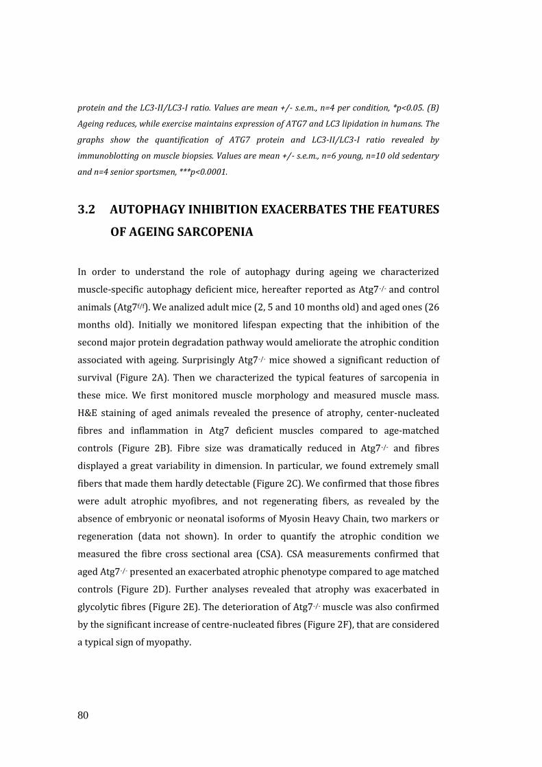
80
protein and the LC3-II/LC3-I ratio. Values are mean +/- s.e.m., n=4 per condition, *p<0.05. (B)
Ageing reduces, while exercise maintains expression of ATG7 and LC3 lipidation in humans. The
graphs show the quantification of ATG7 protein and LC3-II/LC3-I ratio revealed by
immunoblotting on muscle biopsies. Values are mean +/- s.e.m., n=6 young, n=10 old sedentary
and n=4 senior sportsmen, ***p<0.0001.
3.2 AUTOPHAGY INHIBITION EXACERBATES THE FEATURES
OF AGEING SARCOPENIA
In order to understand the role of autophagy during ageing we characterized
muscle-specific autophagy deficient mice, hereafter reported as Atg7-/- and control
animals (Atg7f/f). We analized adult mice (2, 5 and 10 months old) and aged ones (26
months old). Initially we monitored lifespan expecting that the inhibition of the
second major protein degradation pathway would ameliorate the atrophic condition
associated with ageing. Surprisingly Atg7-/- mice showed a significant reduction of
survival (Figure 2A). Then we characterized the typical features of sarcopenia in
these mice. We first monitored muscle morphology and measured muscle mass.
H&E staining of aged animals revealed the presence of atrophy, center-nucleated
fibres and inflammation in Atg7 deficient muscles compared to age-matched
controls (Figure 2B). Fibre size was dramatically reduced in Atg7-/- and fibres
displayed a great variability in dimension. In particular, we found extremely small
fibers that made them hardly detectable (Figure 2C). We confirmed that those fibres
were adult atrophic myofibres, and not regenerating fibers, as revealed by the
absence of embryonic or neonatal isoforms of Myosin Heavy Chain, two markers or
regeneration (data not shown). In order to quantify the atrophic condition we
measured the fibre cross sectional area (CSA). CSA measurements confirmed that
aged Atg7-/- presented an exacerbated atrophic phenotype compared to age matched
controls (Figure 2D). Further analyses revealed that atrophy was exacerbated in
glycolytic fibres (Figure 2E). The deterioration of Atg7-/- muscle was also confirmed
by the significant increase of centre-nucleated fibres (Figure 2F), that are considered
a typical sign of myopathy.
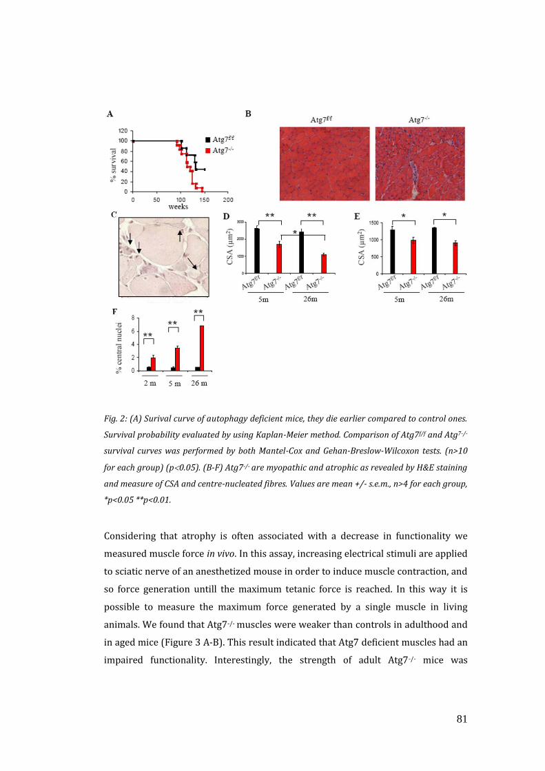
81
Fig. 2: (A) Surival curve of autophagy deficient mice, they die earlier compared to control ones.
Survival probability evaluated by using Kaplan-Meier method. Comparison of Atg7f/f and Atg7-/-
survival curves was performed by both Mantel-Cox and Gehan-Breslow-Wilcoxon tests. (n>10
for each group) (p0.05). (B-F) Atg7-/- are myopathic and atrophic as revealed by H&E staining
and measure of CSA and centre-nucleated fibres. Values are mean +/- s.e.m., n>4 for each group,
*p<0.05 **p<0.01.
Considering that atrophy is often associated with a decrease in functionality we
measured muscle force in vivo. In this assay, increasing electrical stimuli are applied
to sciatic nerve of an anesthetized mouse in order to induce muscle contraction, and
so force generation untill the maximum tetanic force is reached. In this way it is
possible to measure the maximum force generated by a single muscle in living
animals. We found that Atg7-/- muscles were weaker than controls in adulthood and
in aged mice (Figure 3 A-B). This result indicated that Atg7 deficient muscles had an
impaired functionality. Interestingly, the strength of adult Atg7-/- mice was
82
comparable to the force generated by 26 months old controls suggesting an ongoing
precocious ageing. Given that muscle atrophy and weakness are exacerbated in
autophagy-deficient muscles, and that a decline in innervation and loss of motor
units are known to be important endogenous causes of sarcopenia, we tested
whether the ageing-associated muscle loss was due to a possible ongoing
denervation process. We first evaluated the expression pattern of Neural Cell
Adhesion Molecule (N-CAM), that is a well defined re-innervation marker. NCAM
localizes specifically at the neuromuscular junction (NMJ) level in innervated
myofiber, but after denervation it is expressed along the entire fibres. Therefore,
NCAM localization is used as a marker of denervation. As expected, aged control
animals showed some NCAM positive however autophagy-deficient muscles
displayed 5-7 fold more NCAM-positive fibres when compared to age-matched
controls in both adulthood and elderly (Figure 3 C-E).
Fig. 3: (A) Atg7-/- mice show a profound decrease in maximal force generation in adult animals.
Values are mean +/- s.e.m., (n=5), **p<0.01. (B) Ageing reduces force production in both Atg7f/f
and Atg7-/- mice, therefore aggravating the already profound weakness of Atg7-/- mice. Values
are mean +/- s.e.m., n=4, *p<0.05. (C) Representative images of immunostaining for NCAM in
aged mice. (D) Immunoblotting for NCAM protein on muscle extracts from adult and aged GCN
muscles. Atg7-/- muscles express higher levels of NCAM than age-matched Atg7f/f. (E)
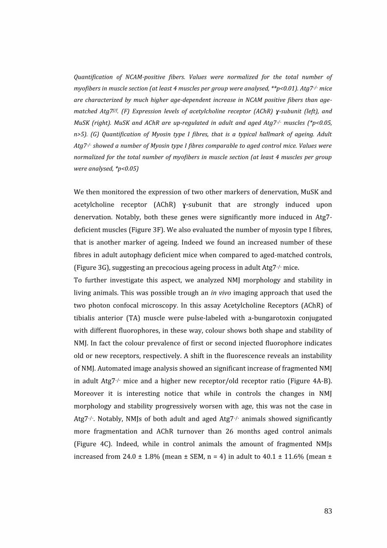
83
Quantification of NCAM-positive fibers. Values were normalized for the total number of
myofibers in muscle section (at least 4 muscles per group were analysed, **p<0.01). Atg7-/- mice
are characterized by much higher age-dependent increase in NCAM positive fibers than age-
matched Atg7f/f. (F) Expression levels of acetylcholine receptor (AChR) ɣ-subunit (left), and
MuSK (right). MuSK and AChR are up-regulated in adult and aged Atg7-/- muscles (*p<0.05,
n>5). (G) Quantification of Myosin type I fibres, that is a typical hallmark of ageing. Adult
Atg7-/- showed a number of Myosin type I fibres comparable to aged control mice. Values were
normalized for the total number of myofibers in muscle section (at least 4 muscles per group
were analysed, *p<0.05)
We then monitored the expression of two other markers of denervation, MuSK and
acetylcholine receptor (AChR) ɣ-subunit that are strongly induced upon
denervation. Notably, both these genes were significantly more induced in Atg7-
deficient muscles (Figure 3F). We also evaluated the number of myosin type I fibres,
that is another marker of ageing. Indeed we found an increased number of these
fibres in adult autophagy deficient mice when compared to aged-matched controls,
(Figure 3G), suggesting an precocious ageing process in adult Atg7-/- mice.
To further investigate this aspect, we analyzed NMJ morphology and stability in
living animals. This was possible trough an in vivo imaging approach that used the
two photon confocal microscopy. In this assay Acetylcholine Receptors (AChR) of
tibialis anterior (TA) muscle were pulse-labeled with a-bungarotoxin conjugated
with different fluorophores, in these way, colour shows both shape and stability of
NMJ. In fact the colour prevalence of first or second injected fluorophore indicates
old or new receptors, respectively. A shift in the fluorescence reveals an instability
of NMJ. Automated image analysis showed an significant increase of fragmented NMJ
in adult Atg7-/- mice and a higher new receptor/old receptor ratio (Figure 4A-B).
Moreover it is interesting notice that while in controls the changes in NMJ
morphology and stability progressively worsen with age, this was not the case in
Atg7-/-. Notably, NMJs of both adult and aged Atg7-/- animals showed significantly
more fragmentation and AChR turnover than 26 months aged control animals
(Figure 4C). Indeed, while in control animals the amount of fragmented NMJs
increased from 24.0 ± 1.8% (mean ± SEM, n = 4) in adult to 40.1 ± 11.6% (mean ±
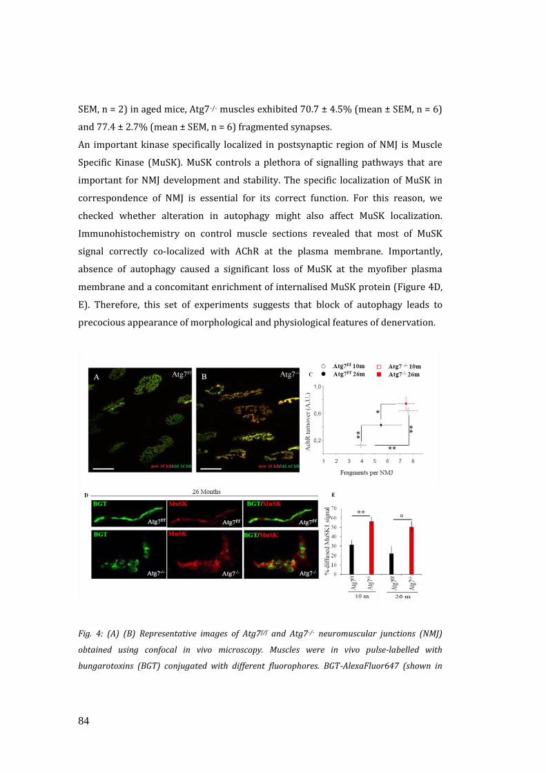
84
SEM, n = 2) in aged mice, Atg7-/- muscles exhibited 70.7 ± 4.5% (mean ± SEM, n = 6)
and 77.4 ± 2.7% (mean ± SEM, n = 6) fragmented synapses.
An important kinase specifically localized in postsynaptic region of NMJ is Muscle
Specific Kinase (MuSK). MuSK controls a plethora of signalling pathways that are
important for NMJ development and stability. The specific localization of MuSK in
correspondence of NMJ is essential for its correct function. For this reason, we
checked whether alteration in autophagy might also affect MuSK localization.
Immunohistochemistry on control muscle sections revealed that most of MuSK
signal correctly co-localized with AChR at the plasma membrane. Importantly,
absence of autophagy caused a significant loss of MuSK at the myofiber plasma
membrane and a concomitant enrichment of internalised MuSK protein (Figure 4D,
E). Therefore, this set of experiments suggests that block of autophagy leads to
precocious appearance of morphological and physiological features of denervation.
Fig. 4: (A) (B) Representative images of Atg7f/f and Atg7-/- neuromuscular junctions (NMJ)
obtained using confocal in vivo microscopy. Muscles were in vivo pulse-labelled with
bungarotoxins (BGT) conjugated with different fluorophores. BGT-AlexaFluor647 (shown in
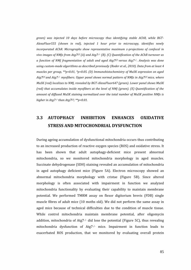
85
green) was injected 10 days before microscopy thus identifying stable AChR, while BGT-
AlexaFluor555 (shown in red), injected 1 hour prior to microscopy, identifies newly
incorporated AChR. Micrographs show representative maximum z-projections of confocal in
vivo images of NMJs from Atg7f/f (A) and Atg7-/- (B). (C) Quantification of the AChR turnover as
a function of NMJ fragmentation of adult and aged Atg7f/f versus Atg7-/-. Analysis was done
using custom-made algorithms as described previously (Roder et al., 2010). Data from at least 4
muscles per group, **p<0.01, *p<0.05. (D) Immunohistochemistry of MuSK expression on aged
Atg7f/f and Atg7-/- myofibers. Upper panel shows normal pattern of NMJs in Atg7f/f mice, where
MuSK (red) localizes to NMJ, revealed by BGT-AlexaFluor647 (green). Lower panel shows MuSK
(red) that accumulates inside myofibers at the level of NMJ (green). (E) Quantification of the
amount of diffused MuSK staining normalized over the total number of MuSK positive NMJs is
higher in Atg7-/- than Atg7f/f; **p<0.01.
3.3 AUTOPHAGY INHIBITION ENHANCES OXIDATIVE
STRESS AND MITOCHONDRIAL DYSFUNCTION
During ageing accumulation of dysfunctional mitochondria occurs thus contributing
to an increased production of reactive oxygen species (ROS) and oxidative stress. It
has been shown that adult autophagy-deficient mice present abnormal
mitochondria, so we monitored mitochondria morphology in aged muscles.
Succinate dehydrogenase (SDH) staining revealed an accumulation of mitochondria
in aged autophagy deficient mice (Figure 5A). Electron microscopy showed an
abnormal mitochondria morphology with cristae (Figure 5B). Since altered
morphology is often associated with impairment in function we analyzed
mitochondria functionality by evaluating their capability to maintain membrane
potential. We performed TMRM assay on flexor digitorium brevis (FDB) single
muscle fibres of adult mice (10 moths old). We did not perform the same assay in
aged mice because of technical difficulties due to the condition of muscle tissue.
While control mitochondria maintain membrane potential, after oligomycin
addition, mitochondria of Atg7-/- did lose the potential (Figure 5C), thus revealing
mitochondria dysfunction of Atg7-/- mice. Impairment in function leads to
exacerbated ROS production, that we monitored by evaluating overall protein
86
carbonylation, that indeed was increased in Atg7-/- mice (Figure 5D). Atg7-/- muscles
showed two-fold increase of carbonylated proteins when compared to age-matched
controls.
Fig. 5: (A) SDH staining on serial sections of aged Atg7f/f and Atg7-/- muscles (TA) shows an
accumulation of abnormal mitochondria in Atg7-/- myofibers. (B) Electron microscopy images
of EDL muscles from aged Atg7f/f and Atg7-/- mice show accumulation of abnormal
mitochondria displaying alterations in size, cristae morphology and matrix density. (C)
Measurement of mitochondrial membrane potential. Isolated FDB muscle fibres from adult
Atg7f/f and Atg7-/- mice were loaded with TMRM, that accumulates in healthy mitochondria that
are able to maintain membrane potential. Atg7-/- mitochondria dissipate membrane potential.
TMRM staining was monitored in at least 20 fibers per group (** p<0.001). (D) Overall protein
carbonylation of aged Atg7f/f and Atg7-/- muscles, revealed by Oxyblot. A representative
immunoblot for carbonylated proteins is depicted on the left, and densitometric quantification
of the carbonylated proteins is in the graph on the right. Aged Atg7-/- mice show higher ongoing
protein carbonylation than Atg7f/f . (n=5, *p<0.05).
Next we performed proteomic approach on carbonylated proteins to determine
which proteins were oxidized in aged control compared to aged Atg7-/- mice. The
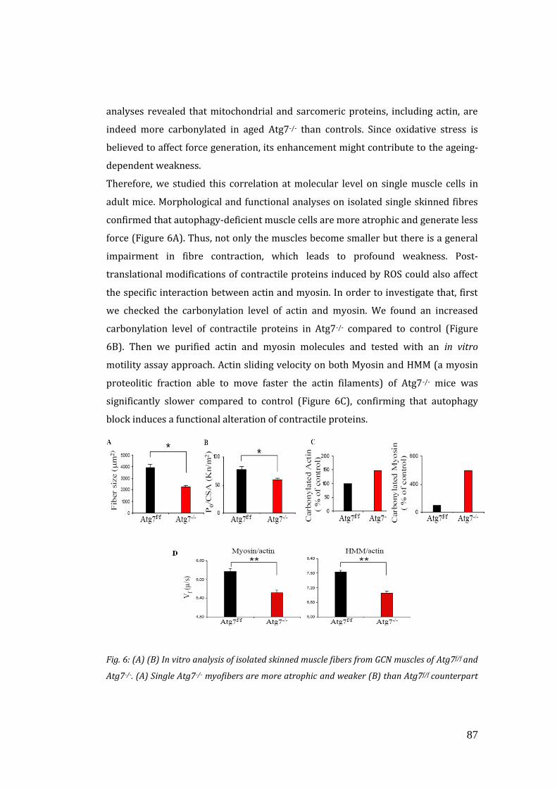
87
analyses revealed that mitochondrial and sarcomeric proteins, including actin, are
indeed more carbonylated in aged Atg7-/- than controls. Since oxidative stress is
believed to affect force generation, its enhancement might contribute to the ageing-
dependent weakness.
Therefore, we studied this correlation at molecular level on single muscle cells in
adult mice. Morphological and functional analyses on isolated single skinned fibres
confirmed that autophagy-deficient muscle cells are more atrophic and generate less
force (Figure 6A). Thus, not only the muscles become smaller but there is a general
impairment in fibre contraction, which leads to profound weakness. Post-
translational modifications of contractile proteins induced by ROS could also affect
the specific interaction between actin and myosin. In order to investigate that, first
we checked the carbonylation level of actin and myosin. We found an increased
carbonylation level of contractile proteins in Atg7-/- compared to control (Figure
6B). Then we purified actin and myosin molecules and tested with an in vitro
motility assay approach. Actin sliding velocity on both Myosin and HMM (a myosin
proteolitic fraction able to move faster the actin filaments) of Atg7-/- mice was
significantly slower compared to control (Figure 6C), confirming that autophagy
block induces a functional alteration of contractile proteins.
Fig. 6: (A) (B) In vitro analysis of isolated skinned muscle fibers from GCN muscles of Atg7f/f and
Atg7-/-. (A) Single Atg7-/- myofibers are more atrophic and weaker (B) than Atg7f/f counterpart
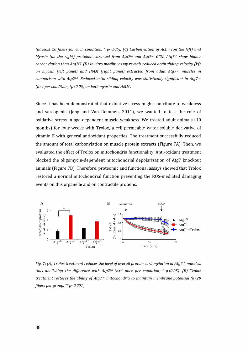
88
(at least 20 fibers for each condition, * p<0.05). (C) Carbonylation of Actin (on the left) and
Myosin (on the right) proteins, extracted from Atg7f/f and Atg7-/- GCN. Atg7-/- show higher
carbonylation than Atg7f/f. (D) In vitro motility assay reveals reduced actin sliding velocity (Vf)
on myosin (left panel) and HMM (right panel) extracted from adult Atg7-/- muscles in
comparison with Atg7f/f. Reduced actin sliding velocity was statistically significant in Atg7-/-
(n=4 per condition, *p<0.05) on both myosin and HMM.
Since it has been demonstrated that oxidative stress might contribute to weakness
and sarcopenia (Jang and Van Remmen, 2011), we wanted to test the role of
oxidative stress in age-dependent muscle weakness. We treated adult animals (10
months) for four weeks with Trolox, a cell-permeable water-soluble derivative of
vitamin E with general antioxidant properties. The treatment successfully reduced
the amount of total carbonylation on muscle protein extracts (Figure 7A). Then, we
evaluated the effect of Trolox on mitochondria functionality. Anti-oxidant treatment
blocked the oligomycin-dependent mitochondrial depolarization of Atg7 knockout
animals (Figure 7B). Therefore, proteomic and functional assays showed that Trolox
restored a normal mitochondrial function preventing the ROS-mediated damaging
events on this organelle and on contractile proteins.
Fig. 7: (A) Trolox treatment reduces the level of overall protein carbonylation in Atg7-/- muscles,
thus abolishing the difference with Atg7f/f (n=4 mice per condition, * p<0.05). (B) Trolox
treatment restores the ability of Atg7-/- mitochondria to maintain membrane potential (n>20
fibers per group, ** p<0.001)
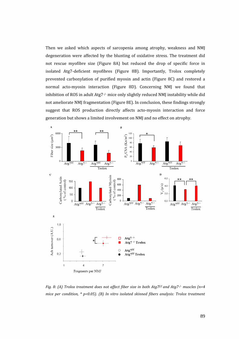
89
Then we asked which aspects of sarcopenia among atrophy, weakness and NMJ
degeneration were affected by the blunting of oxidative stress. The treatment did
not rescue myofibre size (Figure 8A) but reduced the drop of specific force in
isolated Atg7-deficient myofibres (Figure 8B). Importantly, Trolox completely
prevented carbonylation of purified myosin and actin (Figure 8C) and restored a
normal acto-myosin interaction (Figure 8D). Concerning NMJ we found that
inhibition of ROS in adult Atg7-/- mice only slightly reduced NMJ instability while did
not ameliorate NMJ fragmentation (Figure 8E). In conclusion, these findings strongly
suggest that ROS production directly affects acto-myosin interaction and force
generation but shows a limited involvement on NMJ and no effect on atrophy.
Fig. 8: (A) Trolox treatment does not affect fiber size in both Atg7f/f and Atg7-/- muscles (n=4
mice per condition, * p<0.05). (B) In vitro isolated skinned fibers analysis: Trolox treatment
90
rescues the specific muscle force of isolated Atg7-/- myofibers, n=4 mice per condition, * p<0.05.
(C) Trolox treatment reduces the level of actin (left panel) and myosin (right panel) protein
carbonylation, in Atg7-/- muscles, thus abolishing the difference with Atg7f/f n=4 samples. (D)
Trolox treatment rescues actin sliding velocity (Vf) in adult Atg7-/- muscles in in vitro motility
assay, n=4 mice per condition, **p<0.01. (E) Confocal in vivo microscopy: Trolox treatment
slightly reduces AChR turnover (data from at least 4 muscles per group, * p<0.05) but does not
have any effect on NMJ fragmentation of adult Atg7f/f and Atg7-/- mice.
3.4 AUTOPHAGY INHIBITION ALTERS THE RELEASE OF
MUSCLE-DERIVED NEUROTROPHIC FACTORS
Our last findings indicate that even if oxidative stress and ROS generations
contribute to precocious ageing phenotype of Atg7-/- mice, other factors have to be
involved in NMJ degeneration. It is well known that NMJ development and
maintenance is mediated by neurotrophic factors, that are either secreted by the
nerve itself, or either derived from the post-synaptic muscle fibres. So we focused on
the idea that, in our model, autophagy inhibition blocked the release of some factors
that could be important for NMJ. For this reason we screened by qRT-PCR the
expression of several factors that are important for NMJ. Since the changes of NMJ in
autophagy-deficient muscles already occurred in adult mice, we looked for
neurotrophins that were less expressed in both adult and aged Atg7-/-mice. Among
the different neurotrophins only Fibroblast Growth Factor Binding Protein 1
(FGFBP1) was strongly down-regulated in both adult and aged autophagy deficient
mice compared with control counterpart (Figure 9A). Similarly, when we tested the
levels of FGFBP1 in humans we found a significant reduction of its expression in
muscle biopsies of old sedentary subjects (Figure 9B). Importantly, regular exercise
partially counteracted the downregulation of FGFBP1. This data well correlate with
the morphological analyses that showed denervation and atrophy in old sedentary.
These features were strongly attenuated in old sportsmen (Zampieri S et al.
Manuscript in press), which also showed numerous fibre type groupings, a
characteristic sign of re-innervation.
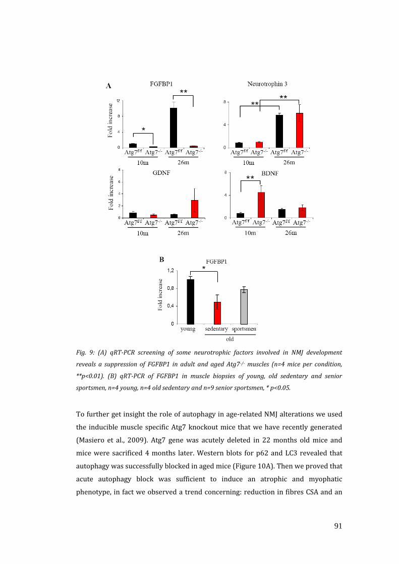
91
Fig. 9: (A) qRT-PCR screening of some neurotrophic factors involved in NMJ development
reveals a suppression of FGFBP1 in adult and aged Atg7-/- muscles (n=4 mice per condition,
**p<0.01). (B) qRT-PCR of FGFBP1 in muscle biopsies of young, old sedentary and senior
sportsmen, n=4 young, n=4 old sedentary and n=9 senior sportsmen, * p<0.05.
To further get insight the role of autophagy in age-related NMJ alterations we used
the inducible muscle specific Atg7 knockout mice that we have recently generated
(Masiero et al., 2009). Atg7 gene was acutely deleted in 22 months old mice and
mice were sacrificed 4 months later. Western blots for p62 and LC3 revealed that
autophagy was successfully blocked in aged mice (Figure 10A). Then we proved that
acute autophagy block was sufficient to induce an atrophic and myophatic
phenotype, in fact we observed a trend concerning: reduction in fibres CSA and an
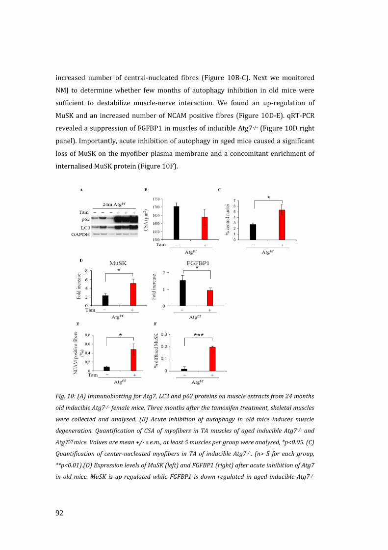
92
increased number of central-nucleated fibres (Figure 10B-C). Next we monitored
NMJ to determine whether few months of autophagy inhibition in old mice were
sufficient to destabilize muscle-nerve interaction. We found an up-regulation of
MuSK and an increased number of NCAM positive fibres (Figure 10D-E). qRT-PCR
revealed a suppression of FGFBP1 in muscles of inducible Atg7-/- (Figure 10D right
panel). Importantly, acute inhibition of autophagy in aged mice caused a significant
loss of MuSK on the myofiber plasma membrane and a concomitant enrichment of
internalised MuSK protein (Figure 10F).
Fig. 10: (A) Immunoblotting for Atg7, LC3 and p62 proteins on muscle extracts from 24 months
old inducible Atg7-/- female mice. Three months after the tamoxifen treatment, skeletal muscles
were collected and analysed. (B) Acute inhibition of autophagy in old mice induces muscle
degeneration. Quantification of CSA of myofibers in TA muscles of aged inducible Atg7-/- and
Atg7f/f mice. Values are mean +/- s.e.m., at least 5 muscles per group were analysed, *p<0.05. (C)
Quantification of center-nucleated myofibers in TA of inducible Atg7-/-. (n> 5 for each group,
**p<0.01).(D) Expression levels of MuSK (left) and FGFBP1 (right) after acute inhibition of Atg7
in old mice. MuSK is up-regulated while FGFBP1 is down-regulated in aged inducible Atg7-/-
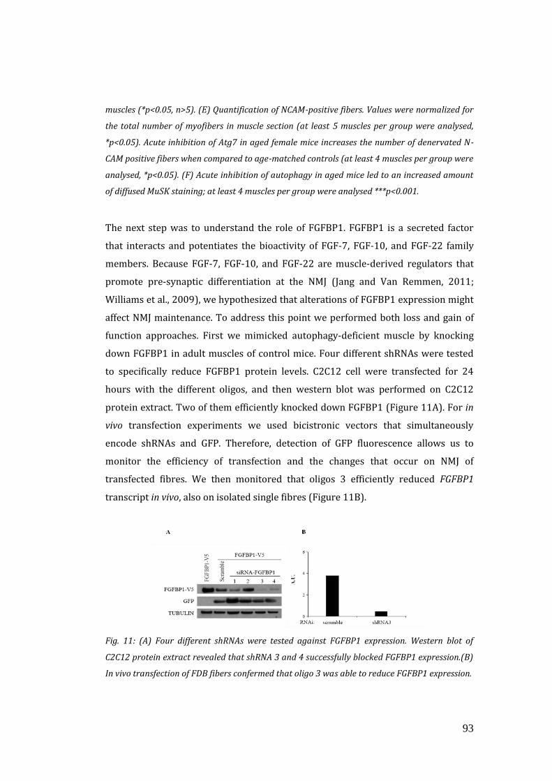
93
muscles (*p<0.05, n>5). (E) Quantification of NCAM-positive fibers. Values were normalized for
the total number of myofibers in muscle section (at least 5 muscles per group were analysed,
*p<0.05). Acute inhibition of Atg7 in aged female mice increases the number of denervated N-
CAM positive fibers when compared to age-matched controls (at least 4 muscles per group were
analysed, *p<0.05). (F) Acute inhibition of autophagy in aged mice led to an increased amount
of diffused MuSK staining; at least 4 muscles per group were analysed ***p<0.001.
The next step was to understand the role of FGFBP1. FGFBP1 is a secreted factor
that interacts and potentiates the bioactivity of FGF-7, FGF-10, and FGF-22 family
members. Because FGF-7, FGF-10, and FGF-22 are muscle-derived regulators that
promote pre-synaptic differentiation at the NMJ (Jang and Van Remmen, 2011;
Williams et al., 2009), we hypothesized that alterations of FGFBP1 expression might
affect NMJ maintenance. To address this point we performed both loss and gain of
function approaches. First we mimicked autophagy-deficient muscle by knocking
down FGFBP1 in adult muscles of control mice. Four different shRNAs were tested
to specifically reduce FGFBP1 protein levels. C2C12 cell were transfected for 24
hours with the different oligos, and then western blot was performed on C2C12
protein extract. Two of them efficiently knocked down FGFBP1 (Figure 11A). For in
vivo transfection experiments we used bicistronic vectors that simultaneously
encode shRNAs and GFP. Therefore, detection of GFP fluorescence allows us to
monitor the efficiency of transfection and the changes that occur on NMJ of
transfected fibres. We then monitored that oligos 3 efficiently reduced FGFBP1
transcript in vivo, also on isolated single fibres (Figure 11B).
Fig. 11: (A) Four different shRNAs were tested against FGFBP1 expression. Western blot of
C2C12 protein extract revealed that shRNA 3 and 4 successfully blocked FGFBP1 expression.(B)
In vivo transfection of FDB fibers confermed that oligo 3 was able to reduce FGFBP1 expression.
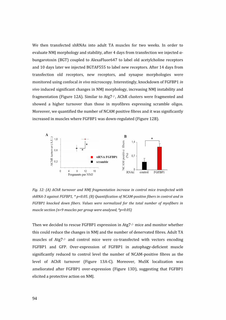
94
We then transfected shRNAs into adult TA muscles for two weeks. In order to
evaluate NMJ morphology and stability, after 4 days from transfection we injected α-
bungarotoxin (BGT) coupled to AlexaFluor647 to label old acetylcholine receptors
and 10 days later we injected BGTAF555 to label new receptors. After 14 days from
transfection old receptors, new receptors, and synapse morphologies were
monitored using confocal in vivo microscopy. Interestingly, knockdown of FGFBP1 in
vivo induced significant changes in NMJ morphology, increasing NMJ instability and
fragmentation (Figure 12A). Similar to Atg7-/-, AChR clusters were fragmented and
showed a higher turnover than those in myofibres expressing scramble oligos.
Moreover, we quantified the number of NCAM positive fibres and it was significantly
increased in muscles where FGFBP1 was down-regulated (Figure 12B).
Fig. 12: (A) AChR turnover and NMJ fragmentation increase in control mice transfected with
shRNA-3 against FGFBP1, * p<0.05. (B) Quantification of NCAM-positive fibers in control and in
FGFBP1 knocked down fibers. Values were normalized for the total number of myofibers in
muscle section (n=9 muscles per group were analysed, *p<0.05)
Then we decided to rescue FGFBP1 expression in Atg7-/- mice and monitor whether
this could reduce the changes in NMJ and the number of denervated fibres. Adult TA
muscles of Atg7-/- and control mice were co-transfected with vectors encoding
FGFBP1 and GFP. Over-expression of FGFBP1 in autophagy-deficient muscle
significantly reduced to control level the number of NCAM-positive fibres as the
level of AChR turnover (Figure 13A-C). Moreover, MuSK localization was
ameliorated after FGFBP1 over-expression (Figure 13D), suggesting that FGFBP1
elicited a protective action on NMJ.
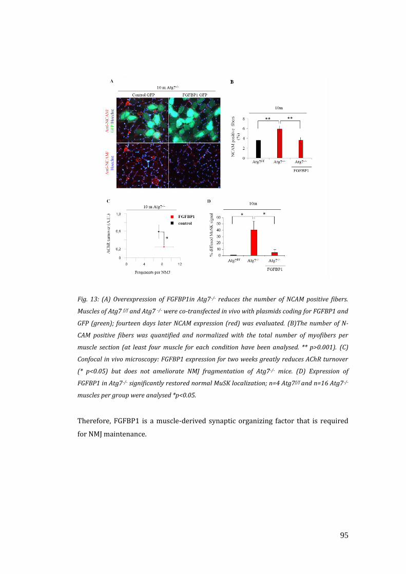
95
Fig. 13: (A) Overexpression of FGFBP1in Atg7-/- reduces the number of NCAM positive fibers.
Muscles of Atg7 f/f and Atg7 -/- were co-transfected in vivo with plasmids coding for FGFBP1 and
GFP (green); fourteen days later NCAM expression (red) was evaluated. (B)The number of N-
CAM positive fibers was quantified and normalized with the total number of myofibers per
muscle section (at least four muscle for each condition have been analysed. ** p>0.001). (C)
Confocal in vivo microscopy: FGFBP1 expression for two weeks greatly reduces AChR turnover
(* p<0.05) but does not ameliorate NMJ fragmentation of Atg7-/- mice. (D) Expression of
FGFBP1 in Atg7-/- significantly restored normal MuSK localization; n=4 Atg7f/f and n=16 Atg7-/-
muscles per group were analysed *p<0.05.
Therefore, FGFBP1 is a muscle-derived synaptic organizing factor that is required
for NMJ maintenance.
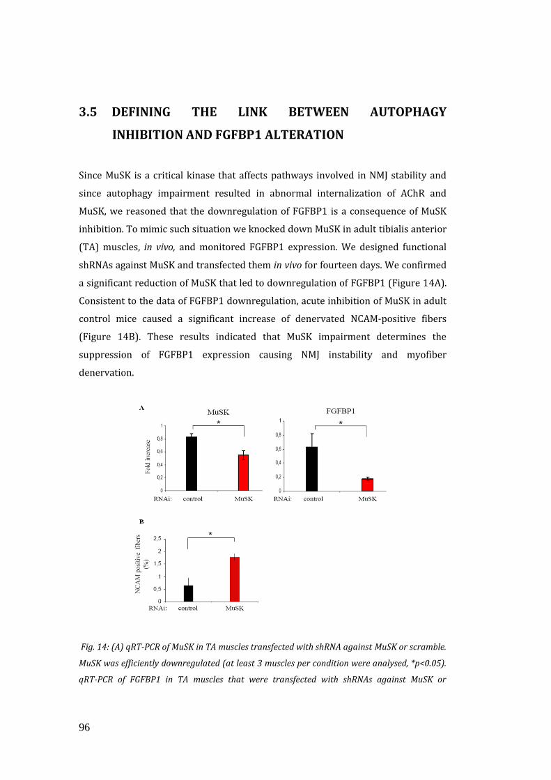
96
3.5 DEFINING THE LINK BETWEEN AUTOPHAGY
INHIBITION AND FGFBP1 ALTERATION
Since MuSK is a critical kinase that affects pathways involved in NMJ stability and
since autophagy impairment resulted in abnormal internalization of AChR and
MuSK, we reasoned that the downregulation of FGFBP1 is a consequence of MuSK
inhibition. To mimic such situation we knocked down MuSK in adult tibialis anterior
(TA) muscles, in vivo, and monitored FGFBP1 expression. We designed functional
shRNAs against MuSK and transfected them in vivo for fourteen days. We confirmed
a significant reduction of MuSK that led to downregulation of FGFBP1 (Figure 14A).
Consistent to the data of FGFBP1 downregulation, acute inhibition of MuSK in adult
control mice caused a significant increase of denervated NCAM-positive fibers
(Figure 14B). These results indicated that MuSK impairment determines the
suppression of FGFBP1 expression causing NMJ instability and myofiber
denervation.
Fig. 14: (A) qRT-PCR of MuSK in TA muscles transfected with shRNA against MuSK or scramble.
MuSK was efficiently downregulated (at least 3 muscles per condition were analysed, *p<0.05).
qRT-PCR of FGFBP1 in TA muscles that were transfected with shRNAs against MuSK or
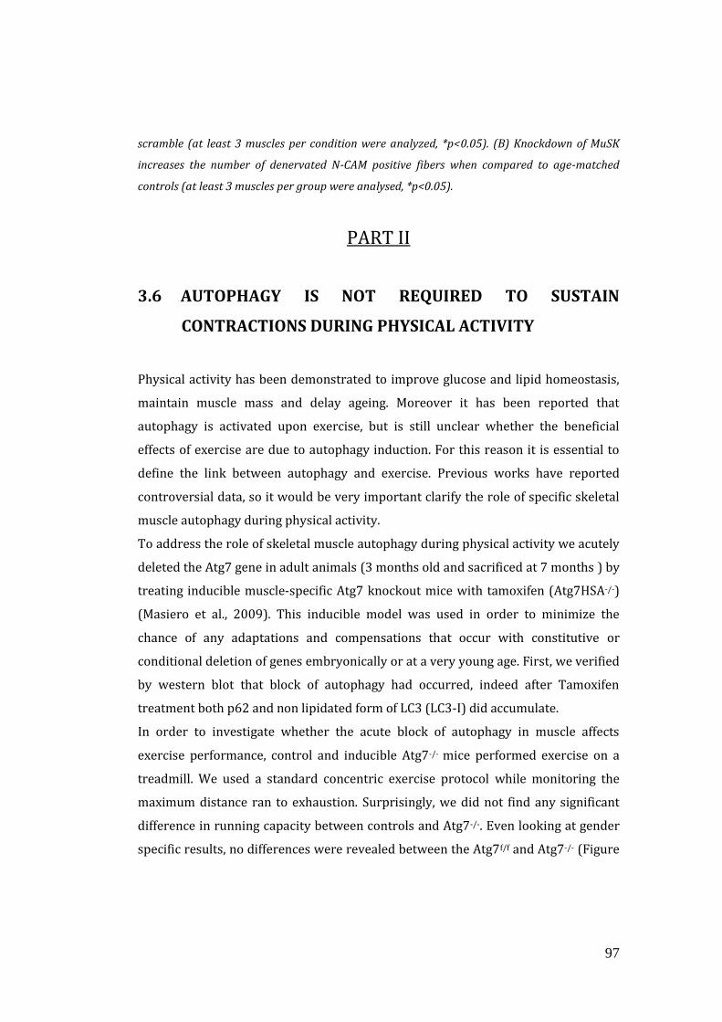
97
scramble (at least 3 muscles per condition were analyzed, *p<0.05). (B) Knockdown of MuSK
increases the number of denervated N-CAM positive fibers when compared to age-matched
controls (at least 3 muscles per group were analysed, *p<0.05).
PART II
3.6 AUTOPHAGY IS NOT REQUIRED TO SUSTAIN
CONTRACTIONS DURING PHYSICAL ACTIVITY
Physical activity has been demonstrated to improve glucose and lipid homeostasis,
maintain muscle mass and delay ageing. Moreover it has been reported that
autophagy is activated upon exercise, but is still unclear whether the beneficial
effects of exercise are due to autophagy induction. For this reason it is essential to
define the link between autophagy and exercise. Previous works have reported
controversial data, so it would be very important clarify the role of specific skeletal
muscle autophagy during physical activity.
To address the role of skeletal muscle autophagy during physical activity we acutely
deleted the Atg7 gene in adult animals (3 months old and sacrificed at 7 months ) by
treating inducible muscle-specific Atg7 knockout mice with tamoxifen (Atg7HSA-/-)
(Masiero et al., 2009). This inducible model was used in order to minimize the
chance of any adaptations and compensations that occur with constitutive or
conditional deletion of genes embryonically or at a very young age. First, we verified
by western blot that block of autophagy had occurred, indeed after Tamoxifen
treatment both p62 and non lipidated form of LC3 (LC3-I) did accumulate.
In order to investigate whether the acute block of autophagy in muscle affects
exercise performance, control and inducible Atg7-/- mice performed exercise on a
treadmill. We used a standard concentric exercise protocol while monitoring the
maximum distance ran to exhaustion. Surprisingly, we did not find any significant
difference in running capacity between controls and Atg7-/-. Even looking at gender
specific results, no differences were revealed between the Atg7f/f and Atg7-/- (Figure
98
15), suggesting that autophagy is not required to sustain muscle contraction during
physical activity.
Fig. 15: (A) Representative western blot analyse, it confirmed p62 and LC3-I accumulation in
Atg7-/- after Tamoxifen treatment. (B) No differences observed in the maximal running distance
during concentric exercise between control and Atg7-/-(at least 10 mice per group were
analysed, *p<0.05); this result was confirmed evaluating also the gender-specific issue, thus
females (C) and males (D).
3.7 AUTOPHAGY IS IMPORTANT TO SUSTAIN PHYSICAL
ACTIVITY THAT PROVOKE DAMAGING CONTRACTIONS
Since autophagy is important for effective protein and organelle turnover as well as
for survival under cellular stress, we tested whether autophagy could have a role in
repairing muscle after damaging contraction. To address this, we performed a
downhill running exercise to induce damaging eccentric contraction in Atg7f/f and
Atg7-/- animals while recording maximal running distance achieved.
We confirmed that autophagy was induced upon exercise in control animals, as
indicated by the lipidation of LC3 and a decrease in p62 in the muscle of Atg7f/f.
Conversely, Atg7-/- maintained their high levels of LC3-I and p62 protein, confirming
the efficient inhibition of autophagy (Figure 16A).
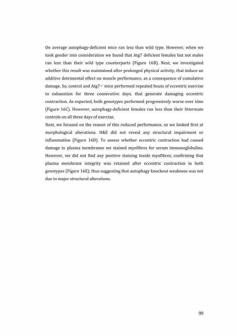
99
On average autophagy-deficient mice ran less than wild type. However, when we
took gender into consideration we found that Atg7 deficient females but not males
ran less than their wild type counterparts (Figure 16B). Next, we investigated
whether this result was maintained after prolonged physical activity, that induce an
additive detrimental effect on muscle performance, as a consequence of cumulative
damage. So, control and Atg7-/- mice performed repeated bouts of eccentric exercise
to exhaustion for three consecutive days, that generate damaging eccentric
contraction. As expected, both genotypes performed progressively worse over time
(Figure 16C). However, autophagy-deficient females ran less than their littermate
controls on all three days of exercise.
Next, we focused on the reason of this reduced performance, so we looked first at
morphological alterations. H&E did not reveal any structural impairment or
inflammation (Figure 16D). To assess whether eccentric contraction had caused
damage to plasma membranes we stained myofibres for serum immunoglobulins.
However, we did not find any positive staining inside myofibres, confirming that
plasma membrane integrity was retained after eccentric contraction in both
genotypes (Figure 16E), thus suggesting that autophagy knockout weakness was not
due to major structural alterations.
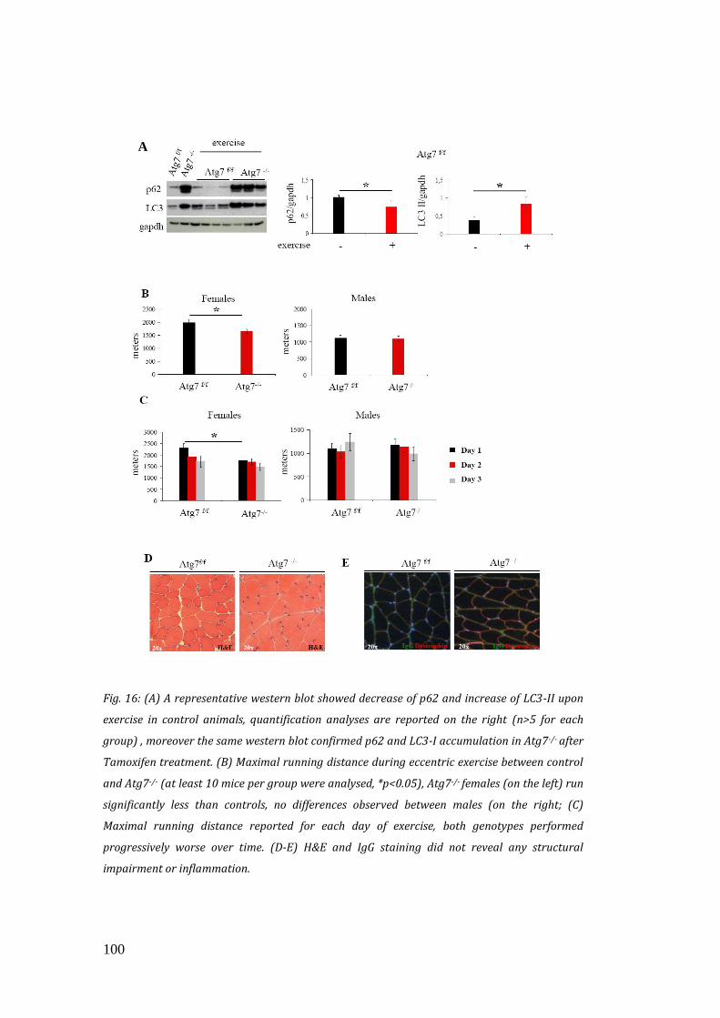
100
Fig. 16: (A) A representative western blot showed decrease of p62 and increase of LC3-II upon
exercise in control animals, quantification analyses are reported on the right (n>5 for each
group) , moreover the same western blot confirmed p62 and LC3-I accumulation in Atg7-/- after
Tamoxifen treatment. (B) Maximal running distance during eccentric exercise between control
and Atg7-/- (at least 10 mice per group were analysed, *p<0.05), Atg7-/- females (on the left) run
significantly less than controls, no differences observed between males (on the right; (C)
Maximal running distance reported for each day of exercise, both genotypes performed
progressively worse over time. (D-E) H&E and IgG staining did not reveal any structural
impairment or inflammation.
A
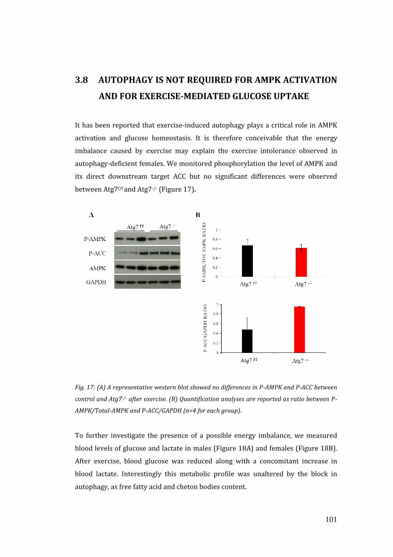
101
3.8 AUTOPHAGY IS NOT REQUIRED FOR AMPK ACTIVATION
AND FOR EXERCISE-MEDIATED GLUCOSE UPTAKE
It has been reported that exercise-induced autophagy plays a critical role in AMPK
activation and glucose homeostasis. It is therefore conceivable that the energy
imbalance caused by exercise may explain the exercise intolerance observed in
autophagy-deficient females. We monitored phosphorylation the level of AMPK and
its direct downstream target ACC but no significant differences were observed
between Atg7f/f and Atg7-/- (Figure 17).
Fig. 17: (A) A representative western blot showed no differences in P-AMPK and P-ACC between
control and Atg7-/- after exercise. (B) Quantification analyses are reported as ratio between P-
AMPK/Total-AMPK and P-ACC/GAPDH (n=4 for each group).
To further investigate the presence of a possible energy imbalance, we measured
blood levels of glucose and lactate in males (Figure 18A) and females (Figure 18B).
After exercise, blood glucose was reduced along with a concomitant increase in
blood lactate. Interestingly this metabolic profile was unaltered by the block in
autophagy, as free fatty acid and cheton bodies content.
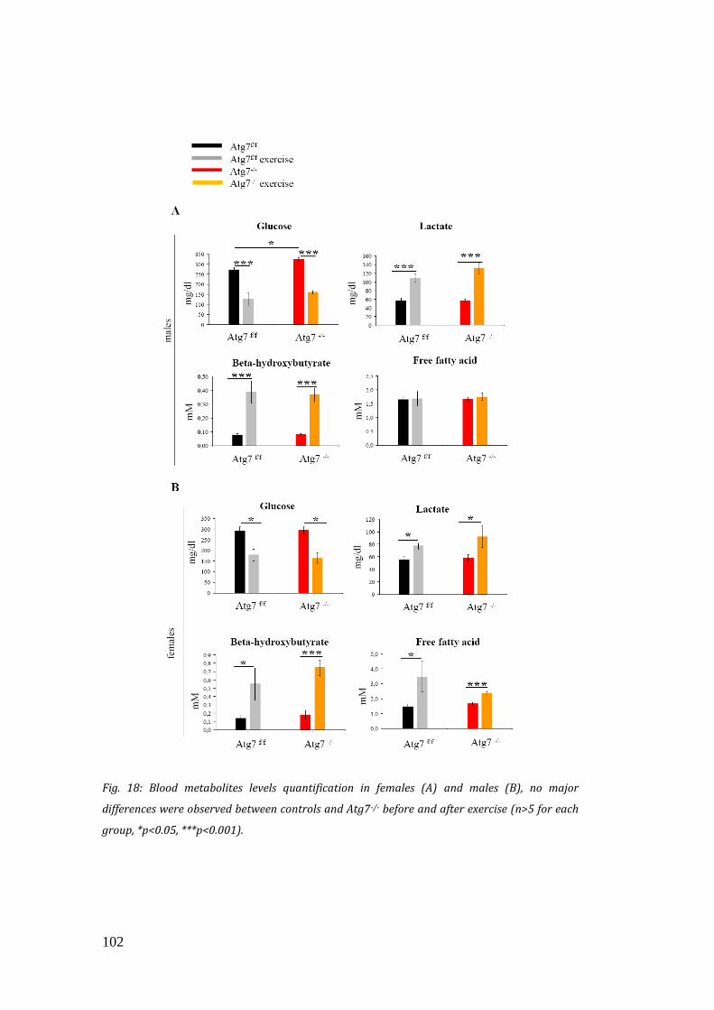
102
Fig. 18: Blood metabolites levels quantification in females (A) and males (B), no major
differences were observed between controls and Atg7-/- before and after exercise (n>5 for each
group, *p<0.05, ***p<0.001).
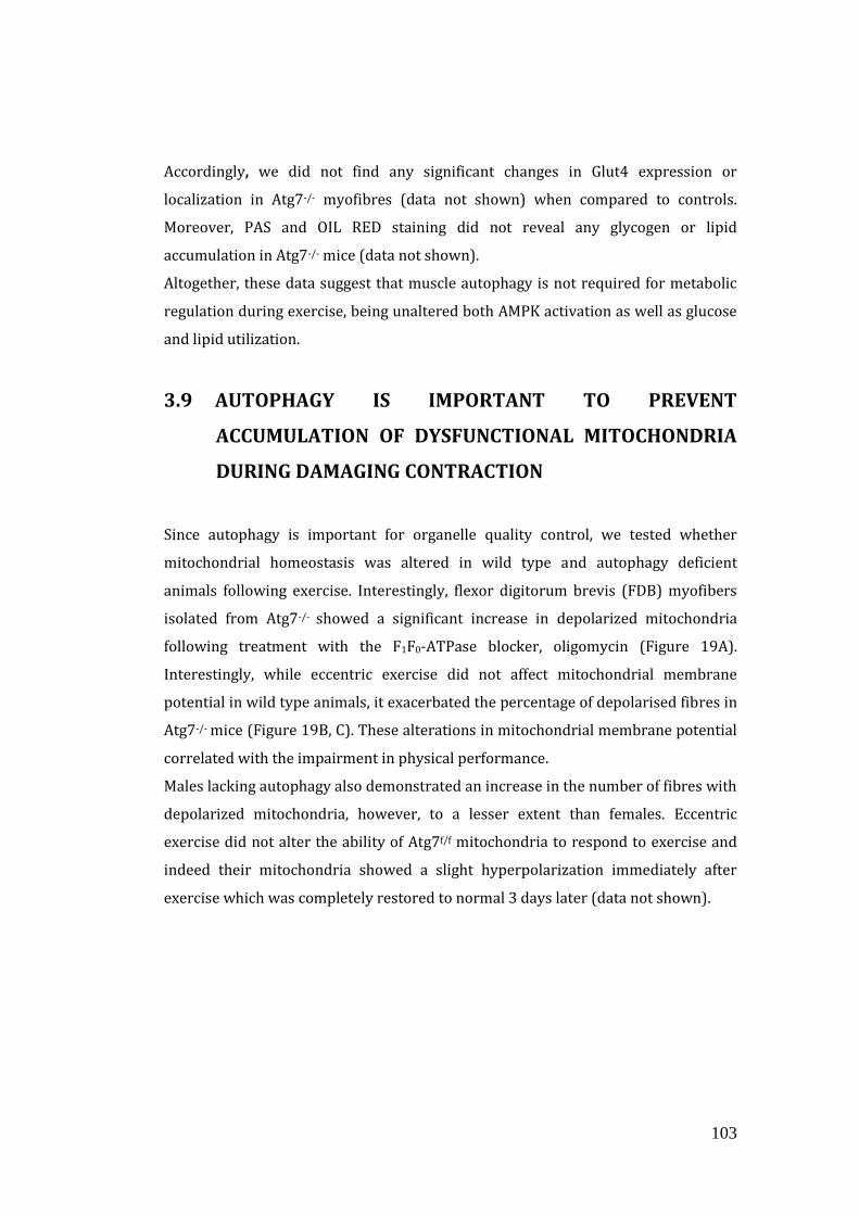
103
Accordingly, we did not find any significant changes in Glut4 expression or
localization in Atg7-/- myofibres (data not shown) when compared to controls.
Moreover, PAS and OIL RED staining did not reveal any glycogen or lipid
accumulation in Atg7-/- mice (data not shown).
Altogether, these data suggest that muscle autophagy is not required for metabolic
regulation during exercise, being unaltered both AMPK activation as well as glucose
and lipid utilization.
3.9 AUTOPHAGY IS IMPORTANT TO PREVENT
ACCUMULATION OF DYSFUNCTIONAL MITOCHONDRIA
DURING DAMAGING CONTRACTION
Since autophagy is important for organelle quality control, we tested whether
mitochondrial homeostasis was altered in wild type and autophagy deficient
animals following exercise. Interestingly, flexor digitorum brevis (FDB) myofibers
isolated from Atg7-/- showed a significant increase in depolarized mitochondria
following treatment with the F1F0-ATPase blocker, oligomycin (Figure 19A).
Interestingly, while eccentric exercise did not affect mitochondrial membrane
potential in wild type animals, it exacerbated the percentage of depolarised fibres in
Atg7-/- mice (Figure 19B, C). These alterations in mitochondrial membrane potential
correlated with the impairment in physical performance.
Males lacking autophagy also demonstrated an increase in the number of fibres with
depolarized mitochondria, however, to a lesser extent than females. Eccentric
exercise did not alter the ability of Atg7f/f mitochondria to respond to exercise and
indeed their mitochondria showed a slight hyperpolarization immediately after
exercise which was completely restored to normal 3 days later (data not shown).
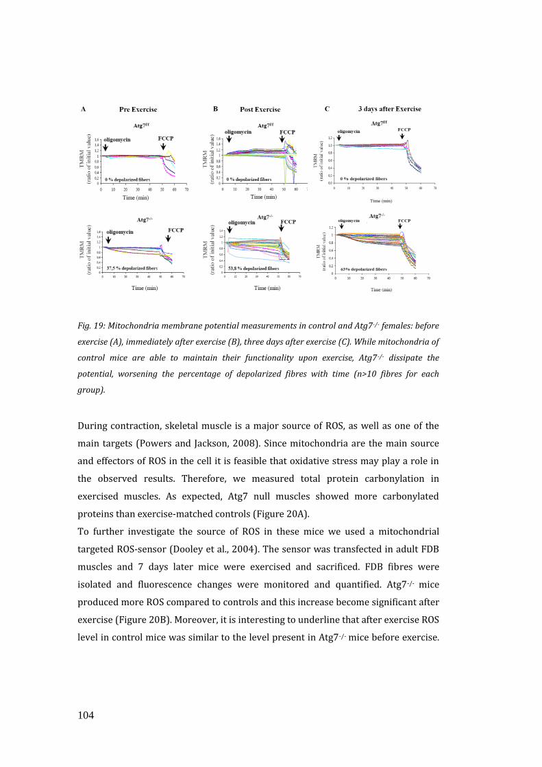
104
Fig. 19: Mitochondria membrane potential measurements in control and Atg7-/- females: before
exercise (A), immediately after exercise (B), three days after exercise (C). While mitochondria of
control mice are able to maintain their functionality upon exercise, Atg7 -/- dissipate the
potential, worsening the percentage of depolarized fibres with time (n>10 fibres for each
group).
During contraction, skeletal muscle is a major source of ROS, as well as one of the
main targets (Powers and Jackson, 2008). Since mitochondria are the main source
and effectors of ROS in the cell it is feasible that oxidative stress may play a role in
the observed results. Therefore, we measured total protein carbonylation in
exercised muscles. As expected, Atg7 null muscles showed more carbonylated
proteins than exercise-matched controls (Figure 20A).
To further investigate the source of ROS in these mice we used a mitochondrial
targeted ROS-sensor (Dooley et al., 2004). The sensor was transfected in adult FDB
muscles and 7 days later mice were exercised and sacrificed. FDB fibres were
isolated and fluorescence changes were monitored and quantified. Atg7-/- mice
produced more ROS compared to controls and this increase become significant after
exercise (Figure 20B). Moreover, it is interesting to underline that after exercise ROS
level in control mice was similar to the level present in Atg7-/- mice before exercise.
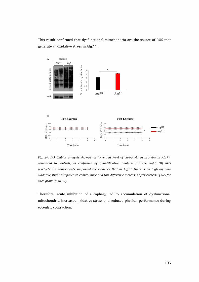
105
This result confirmed that dysfunctional mitochondria are the source of ROS that
generate an oxidative stress in Atg7-/-.
Fig. 20: (A) Oxiblot analysis showed an increased level of carbonylated proteins in Atg7-/-
compared to controls, as confirmed by quantification analyses (on the right. (B) ROS
production measurements supported the evidence that in Atg7-/- there is an high ongoing
oxidative stress compared to control mice and this difference increases after exercise. (n=5 for
each group *p<0.05).
Therefore, acute inhibition of autophagy led to accumulation of dysfunctional
mitochondria, increased oxidative stress and reduced physical performance during
eccentric contraction.
106
3.10 ANTI-OXIDANT TREATMENT DID NOT AMELIORATE
THE PHYSICAL PERFORMANCE OF ATG7 KNOCKOUT
BUT BLOCKED AUTOPHAGY IN CONTROLS WORSENING
MITOCHONDRIAL FUNCTION AND RUNNING CAPACITY
Excessive oxidative stress has been documented to impair muscle function, which
could potentially explain the reduced physical performance of Atg7-/- mice.
(Peternelj and Coombes, 2011; Powers and Jackson, 2008).
In order to evaluate whether reducing oxidative stress could ameliorate their
physical performance, we treated female Atg7-/- and control mice with the
antioxidant N-Acetyl-Cysteine (NAC) for 6 weeks. and then subjected them to
eccentric exercise. Surprisingly, NAC treatment severely impaired performance of
control but did not elicit any benefit in inducible Atg7-/- mice (Figure 21). Indeed, the
difference in the running distance between control and Atg7-/- mice was abolished at
both one and three days of exercise.
Fig. 21: The maximal running distance of females control mice decreased after NAC treatment
from the first day (on the left) till the end (on the right) of the protocol. No difference between
maximal running distance between control and Atg7-/- mice (n>5 for each group *p<0.05,
***p<0.001).
We first checked whether NAC treatment affected general protein carbonylation.
The level of carbonylated proteins was reduced both in control and Atg7-/- mice after
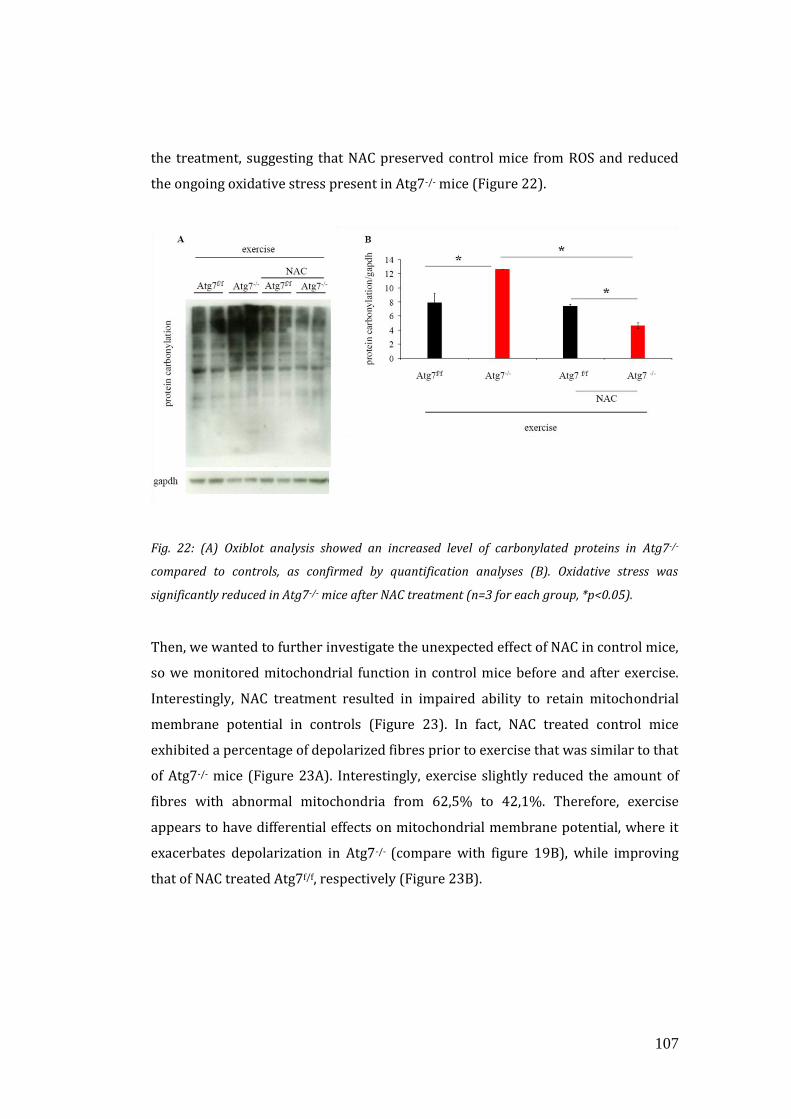
107
the treatment, suggesting that NAC preserved control mice from ROS and reduced
the ongoing oxidative stress present in Atg7-/- mice (Figure 22).
Fig. 22: (A) Oxiblot analysis showed an increased level of carbonylated proteins in Atg7-/-
compared to controls, as confirmed by quantification analyses (B). Oxidative stress was
significantly reduced in Atg7-/- mice after NAC treatment (n=3 for each group, *p<0.05).
Then, we wanted to further investigate the unexpected effect of NAC in control mice,
so we monitored mitochondrial function in control mice before and after exercise.
Interestingly, NAC treatment resulted in impaired ability to retain mitochondrial
membrane potential in controls (Figure 23). In fact, NAC treated control mice
exhibited a percentage of depolarized fibres prior to exercise that was similar to that
of Atg7-/- mice (Figure 23A). Interestingly, exercise slightly reduced the amount of
fibres with abnormal mitochondria from 62,5% to 42,1%. Therefore, exercise
appears to have differential effects on mitochondrial membrane potential, where it
exacerbates depolarization in Atg7-/- (compare with figure 19B), while improving
that of NAC treated Atg7f/f, respectively (Figure 23B).
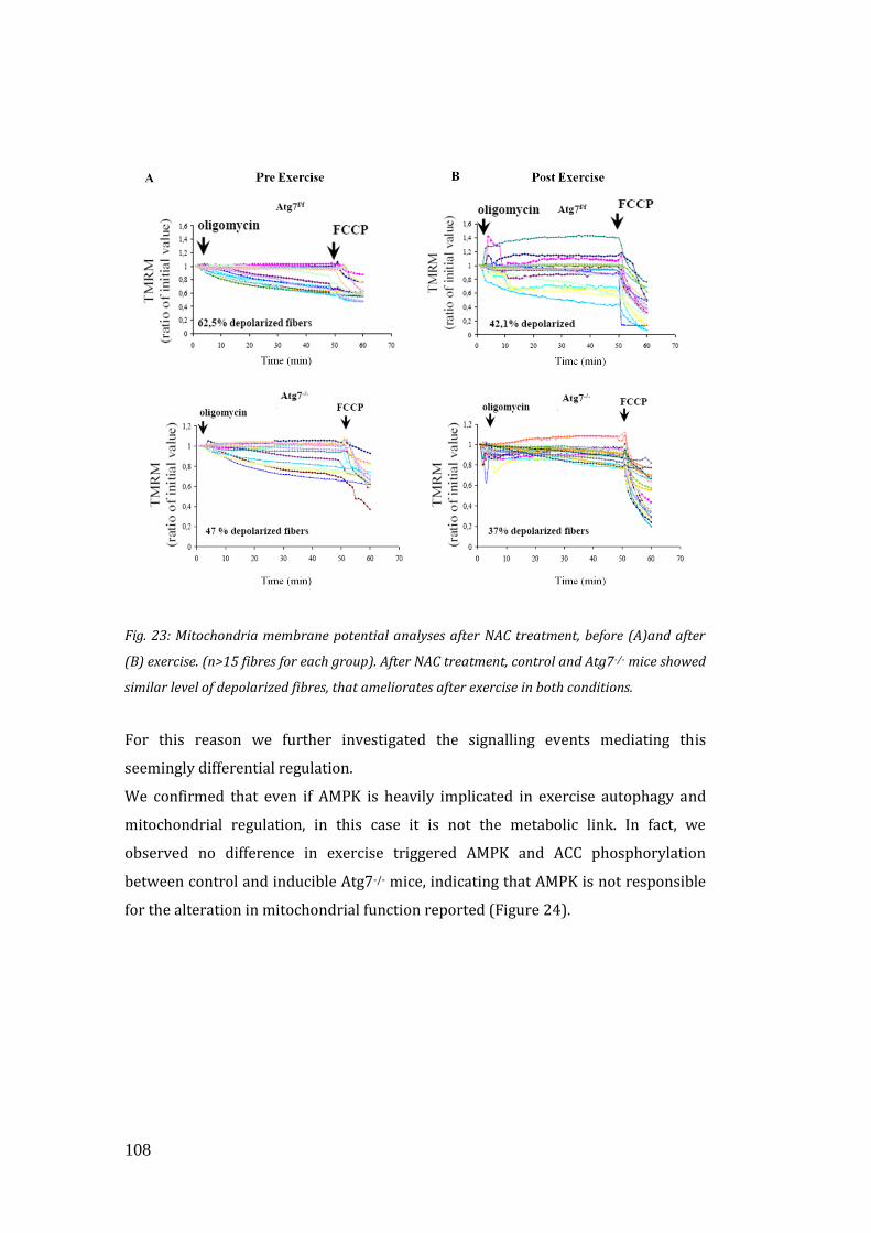
108
Fig. 23: Mitochondria membrane potential analyses after NAC treatment, before (A)and after
(B) exercise. (n>15 fibres for each group). After NAC treatment, control and Atg7-/- mice showed
similar level of depolarized fibres, that ameliorates after exercise in both conditions.
For this reason we further investigated the signalling events mediating this
seemingly differential regulation.
We confirmed that even if AMPK is heavily implicated in exercise autophagy and
mitochondrial regulation, in this case it is not the metabolic link. In fact, we
observed no difference in exercise triggered AMPK and ACC phosphorylation
between control and inducible Atg7-/- mice, indicating that AMPK is not responsible
for the alteration in mitochondrial function reported (Figure 24).
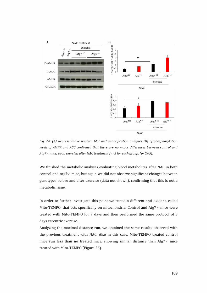
109
Fig. 24: (A) Representative western blot and quantification analyses (B) of phosphorylation
levels of AMPK and ACC confirmed that there are no major differences between control and
Atg7-/- mice, upon exercise, after NAC treatment (n=5 for each group, *p<0.05).
We finished the metabolic analyses evaluating blood metabolites after NAC in both
control and Atg7-/- mice, but again we did not observe significant changes between
genotypes before and after exercise (data not shown), confirming that this is not a
metabolic issue.
In order to further investigate this point we tested a different anti-oxidant, called
Mito-TEMPO, that acts specifically on mitochondria. Control and Atg7-/- mice were
treated with Mito-TEMPO for 7 days and then performed the same protocol of 3
days eccentric exercise.
Analyzing the maximal distance run, we obtained the same results observed with
the previous treatment with NAC. Also in this case, Mito-TEMPO treated control
mice run less than no treated mice, showing similar distance than Atg7-/- mice
treated with Mito-TEMPO (Figure 25).
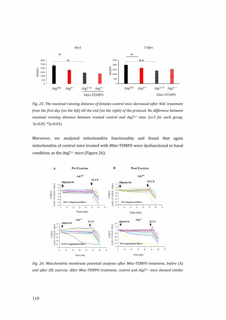
110
Fig. 25: The maximal running distance of females control mice decreased after NAC treatment
from the first day (on the left) till the end (on the right) of the protocol. No difference between
maximal running distance between treated control and Atg7-/- mice (n>3 for each group,
*p<0.05, **p<0.01).
Moreover, we analyzed mitochondria functionality and found that again
mitochondria of control mice treated with Mito-TEMPO were dysfunctional in basal
condition, as the Atg7-/- mice (Figure 26).
Fig. 26: Mitochondria membrane potential analyses after Mito-TEMPO treatment, before (A)
and after (B) exercise. After Mito-TEMPO treatment, control and Atg7-/- mice showed similar
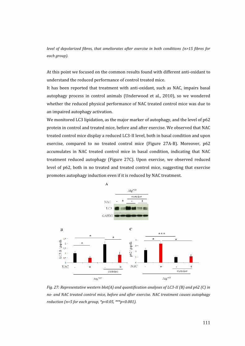
111
level of depolarized fibres, that ameliorates after exercise in both conditions (n>15 fibres for
each group).
At this point we focused on the common results found with different anti-oxidant to
understand the reduced performance of control treated mice.
It has been reported that treatment with anti-oxidant, such as NAC, impairs basal
autophagy process in control animals (Underwood et al., 2010), so we wondered
whether the reduced physical performance of NAC treated control mice was due to
an impaired autophagy activation.
We monitored LC3 lipidation, as the major marker of autophagy, and the level of p62
protein in control and treated mice, before and after exercise. We observed that NAC
treated control mice display a reduced LC3-II level, both in basal condition and upon
exercise, compared to no treated control mice (Figure 27A-B). Moreover, p62
accumulates in NAC treated control mice in basal condition, indicating that NAC
treatment reduced autophagy (Figure 27C). Upon exercise, we observed reduced
level of p62, both in no treated and treated control mice, suggesting that exercise
promotes autophagy induction even if it is reduced by NAC treatment.
Fig. 27: Representative western blot(A) and quantification analyses of LC3-II (B) and p62 (C) in
no- and NAC treated control mice, before and after exercise. NAC treatment causes autophagy
reduction (n=5 for each group, *p<0.05, ***p<0.001).
112
These findings suggest that impaired physical performance of NAC treated control
mice was due to a reduction in autophagy process, mediated by NAC treatment.
Altogether, these findings underline the critical role of autophagy in the
maintenance of proper mitochondrial function. Moreover, we highlight an important
physiological role of oxidative stress for basal autophagy regulation in skeletal
muscle.
113
4. DISCUSSION
In this work, by using muscle specific autophagy-deficient (Atg7-/-), we investigated
the role of autophagy in skeletal muscle during ageing and exercise. We analyzed
how decreased autophagy is related to ageing and the reason why autophagy
reactivation can lead to ameliorated ageing features and increased lifespan.
PART I
In the first part of the work, we studied the role of autophagy during ageing in
muscle tissue. Our results highlight two important functions of autophagy in
sarcopenia, in fact it is responsible for the removal of damaged mitochondria that
produce ROS, and for the signalling that induces the release of the neurotrophic
factor FGFBP1 to maintain NMJ and muscle–nerve interaction.
Ageing leads to the functional degeneration of tissues and this is mainly due to
accumulation of damaged DNA, proteins and organelles. Autophagy is a catabolic
process essential for the maintenance of tissue homeostasis. It is responsible for the
removal of dysfunctional proteins that are prone to aggregate, and for proper
organelle turnover, such as mitochondria, in order to reduce ROS production and
preserve DNA stability (Sandri, 2010).
Due to these actions and to the fact that autophagy declines with age, there is
consensus in considering autophagy as an anti- ageing system. Indeed, genetic
evidences in flies and worms sustain this concept but this indication is still lacking in
mammals (Lee et al., 2010b; Rubinsztein et al., 2011; Simonsen et al., 2008; Madeo
et al., 2010).
For what concerns mammals, there is a more complicated scenario, because
autophagy knockout mice die at newborn stage due to a general energy failure
(Kuma et al., 2004) and most of the tissue specific knockouts (e.g. in brain or
heart)(Hara et al., 2006; Komatsu et al., 2006; Nakai et al., 2007) show severe organ
114
dysfunctions that precludes ageing studies. For this reason the link between
autophagy and ageing is still lacking in mammals.
Our conditional model of muscle-specific autophagy deficiency allowed us to analyse
the role of autophagy in muscles of adult and aged animal. Our findings support the
notion that autophagy failure contributes to ageing. Adult Atg7-/- mice are
characterized by mitochondrial dysfunction, oxidative stress, weakness, atrophy and
loss of innervations; those are typical features of the ageing state, thus suggesting
that autophagy-deficiency leads to premature ageing. Moreover, we found that
altogether these events did shorten the lifespan of autophagy-deficient animals.
Ageing is a multisystemic disorder that affects multiple organs leading to alterations
of metabolism and tissue function. The post-mitotic tissues such as brain, heart and
skeletal muscle are the most susceptible to age-related diseases and precocious
ageing when some dysfunction occurs. Interestingly, among the different age-related
features, only one is conserved in several species, such as C.elegans, Drosophila, and
mammals, including humans, that is the loss of muscle force. Therefore, muscle is a
proper tissue that can be used to investigate ageing mechanisms. Moreover, muscles
play an important role in the regulation of glucose, lipids and therefore in the
general control of metabolism. Recent studies in Drosophila sustained the important
role of muscle in healthy ageing. It has been shown that the over-expression of FoxO
transcription factor, (that leads to autophagy activation), in the muscles of flies,
preserves muscle function and increases longevity (Demontis and Perrimon, 2010),
decreasing the accumulation of protein aggregates also in other tissues. On the
contrary the over-expression of FoxO in adipose tissue, did not induce any changes.
These results confirm that maintenance of a normal autophagy level in skeletal
muscle, is sufficient to induce positively effects on whole-body metabolism,
suggesting an important regulatory role for muscle tissue (Demontis and Perrimon,
2010).
Muscle-specific autophagy deficient mice allowed us to dissect the role of autophagy
during ageing. Our findings support the notion that autophagy is critical to prevent
sarcopenia. Indeed autophagy inhibition is sufficient to trigger a precocious ageing
state in adult mice. It has been shown that oxidative stress increases during ageing,
because of a reduction in autophagy flux and accumulation of dysfunctional
115
mitochondria. Since mitochondria are the main producers of ROS, dysfunctional
mitochondria are thought to play a key role in decline of muscle function. At old age,
a significant proportion of the mitochondria are abnormally enlarged, more rounded
in shape and display vacuolization in the matrix and shorter christae (Peterson et al.,
2012). We found that block of autophagy leads to accumulation of mitochondria
with altered morphology and function. Accordingly, in the previous work it was
showed that energy-stress sensor, AMPK, is activated in Atg7-/- muscles (Masiero et
al., 2009). Importantly, we showed that mitochondrial dysfunction can be reversed
by inhibiting ROS production through the anti-oxidant Trolox. This finding is
consistent with a recent report that showed amelioration of age-related deficit,
including mitochondrial function, by expressing a mitochondrial-targeted human
catalase (Lee et al., 2010a). An higher production of ROS, leads to increased protein
carbonylation, that mainly affects mitochondrial and structural proteins, such as
actin and myosin. This contributes to impaired muscle contraction and weakness,
caused by an altered actin/myosin interaction and a reduction in force generation.
Trolox treatment reverted both protein oxidation and actin sliding properties,
ultimately ameliorating muscle force. It is well established that changes in muscle
mass and strength tend to be dissociated in elderly persons, being the decline of
muscle strength three times faster than the decrease of muscle mass (Peterson et al.,
2012). This notion suggests that alteration in the quality of contractile proteins
plays critical role during age-related decrease of muscle force. Importantly, while
anti-oxidant ameliorates acto/myosin functionality, it does not protect from muscle
atrophy. However we cannot exclude that extending Trolox treatment would
counteract muscle atrophy as well. Autophagy impairment also causes instability
and degeneration of NMJ. Previous studies reported that mitochondrial ROS
production may contribute to NMJ instability (Dobrowolny et al., 2008; Jang et al.,
2012; Jang et al., 2010). But in our hands anti-oxidant treatment only slightly
ameliorated NMJ stability of autophagy deficient mice, with no effect on
fragmentation suggesting that other players were necessarily involved in NMJ
maintenance. Indeed muscle tissue itself plays an important role in the signalling for
NMJ development and maintenance (Johnson-Venkatesh and Umemori, 2010). In
particular, molecules are secreted retrogradely from muscle to nerve, thus affecting
116
motor neuron survival, growth and maintenance (Dobrowolny et al., 2005;
Funakoshi et al., 1995). We found that FGFBP1 is required for NMJ maintenance and
that a normal autophagic flux is critical for its correct regulation. Knocking down
FGFBP1 expression for 14 days in control muscles was sufficient to trigger
fragmentation of NMJs and to increase AChR turnover. Moreover, these effects
appear after only 2 weeks of FGFBP1 inhibition. Prolonged FGFBP1 perturbation
might even worsen NMJ instability and fragmentation. In the opposite situation,
when we over-expressed FGFBP1 in Atg7-/- mice we observed an ameliorated NMJ
stability and rescue in the number of NCAM positive fibres, thus confirming that
FGFBP1 is a muscle-derived synaptic organizing factor that is required for NMJ
maintenance.
The alteration of FGFBP1 expression is a consequence of the impairment of
endocytic trafficking that impacts on AChR cluster and MuSK activity on
downstream signalling. In fact surface expression of MuSK, a kinase that affects a
plethora of signalling pathways that are important for NMJ stability, is regulated by
the endocytic pathway (Punga and Ruegg, 2012). MuSK does not properly localize at
the NMJ in Atg7 knockout, being internalized instead of being at the surface of the
myofibre. However, it was re-localized at the NMJ when FGFBP1 was over-expressed
in Atg7-/- mice.
Our data strongly support the concept that autophagy, in a post-mitotic tissue, is
required for healthy ageing being critical for the correct interplay between muscle
and nerve and for the quality-control of mitochondria. Moreover we have evidences
to believe that the beneficial effects of caloric restriction and exercise on ageing are
a consequence of autophagy reactivation. Future works will investigate the link
between autophagy inhibition, altered expression of FGFBP1 and MuSK.
117
PART II
In the second part of the work, we investigated the connection between autophagy
and physical exercise.
We found that autophagy is required for healthy ageing. Lifestyle adaptations, in
particular caloric restriction and physical activity contributes to increase lifespan
ameliorating some ageing features (Melov et al., 2007; Fontana et al., 2010; Sandri et
al., 2013). In particular, we analyzed human biopsies of young, aged sedentary
subjects or senior sportsmen. We confirmed that autophagy is reduced with ageing,
but upon exercise it is reactivated also in elderly. So it is important to understand
the role of autophagy during physical activity, and to investigate whether the
beneficial effects of exercise (that are also visible in aged people) are directly due to
autophagy reactivation.
Autophagy is activated upon exercise in several tissues, in particular it has been
shown that acute bout of exercise activates autophagosome formation in skeletal
muscles (Grumati et al., 2010; Grumati et al., 2011a; Grumati et al., 2011b; He et al.,
2012; Rubinsztein et al., 2011; Wohlgemuth et al., 2010). It has previously reported
that autophagy is required for proper energy provision and glucose homeostasis
during muscle contraction potentially through AMPK activation and its downstream
targets (He et al., 2012). However, another study reached the opposite conclusion
indicating that autophagy inhibition results in an improved blood glucose profile
(Kim et al., 2013). Therefore, up to now there is no general consensus about the
metabolic impact of autophagy during exercise, whether this effect is cell-
autonomous and the mechanism underlying has not been fully understood yet. In
order to address this issue we used inducible muscle-specific autophagy knockout
mice, that were recently generated in our laboratory (Masiero et al., 2009). In these
mice a muscle-specific acute deletion of the Atg7 gene was induced upon Tamoxifen
treatment just before exercise. We used this model to avoid any possible
compensation mechanism, that occur in conditional models.
We observed that block of autophagy in skeletal muscle did not impact on physical
performance, glucose homeostasis or AMPK activation in our mice. Accordingly, a
recent report where heterozygous mice for BECLIN1 were subjected to exercise also
118
found no differences in running capacity, providing further evidence that autophagy
is not required during an acute bout of exercise (Safdar et al., 2011). The same study
also demonstrated that the absence of exercise-induced autophagy results in a lack
of exercise training-induced adaptations; thus suggesting that autophagy is required
for muscle adaptations to chronic exercise which need mitochondrial turnover and
remodelling (Safdar et al., 2011). So we wondered whether reduced performance
was caused by impaired mitochondria functionality. Our data, indeed, support the
idea that autophagy is critical for removing dysfunctional mitochondria during
damaging contraction, and in fact the absence of autophagy leads to reduced
running performance mainly due to increased ROS production and accumulation of
dysfunctional mitochondria. Interestingly, this effect is gender specific being females
more affected than males. These data are in line with the concept that females are
more prone to muscle wasting during catabolic conditions. Moreover, one possibility
could be that impairment in protein and organelle turnover might manifest more
rapidly in females than in males, as it is known that in different tissues, such as
brown adipose tissue, or mechanisms, such as oxidative stress response and
mitochondria biogenesis, there are sex-dependent differences. (Nadal-casellas et al.,
2013; Huges and Hekimi, 2011). Nevertheless it is plausible that different exercise
regimens or a more chronic autophagy inhibition may unravel alterations in muscle
performance in males as well. Further research into gender difference is required in
order to discern the mechanisms responsible for male protection and female
predisposition.
We wondered then whether reduction of oxidative stress could ameliorate physical
performance of Atg7-/- mice. The chronic use of the antioxidant N-acetyl-cysteine did
not ameliorate Atg7-/- performance, but on the contrary impaired the one of control
animals. Moreover control mice treated with NAC showed a decreased basal and
exercise-induced autophagy level. This result suggest that a basal level of ROS is
necessary as signal to maintain redox homeostasis and autophagy activation
mechanisms. Other works reported that ‘physiological ROS’ are important to
mediate autophagy activation, thus maintaining tissue homeostasis (Underwood et
al., 2010; Owusu-Ansah et al., 2013). Moreover, our findings correlate with the
notion that anti-oxidants are detrimental for exercise-induced benefits, that has
119
already been reported in humans, although the mechanisms remain unclear (Ristow
et al., 2011). We did not found significant changes in AMPK activation nor in blood
metabolites that could explain the reduced physical performance. Moreover these
data were confirmed when we acutely treated mice with a mitochondria-specific
antioxidant.
Since we did not observed metabolic changes, we thought that one possible
explanation could involve mitochondria-derived metabolites, which play an
important role in signalling and could therefore impinge on cellular adaptations to
stress. In our case, dysfunctional mitochondria can release factors that damage
muscle tissue or, on the contrary, mitochondria impairment can lead to lack of
certain important secreted factors. For instance, alpha-ketoglutarate-derived
glutamine has been found to inhibit mTOR activity and activate autophagy (van der
Vos et al., 2012). Importantly a recent work reported that impaired mitochondrial
function in muscle specific Atg7 knockouts is protective from diet-induced obesity
due to the induction of a mitochondrial stress mitokine, FGF21 (Kim et al., 2013).
Therefore, it is possible that dysfunctional mitochondria generate metabolites or
myokines that are involved in pathways important for myofibres function during
exhausting and damaging muscle contraction. Moreover, the detrimental effects of
anti-oxidants on physical performance underline the important role of mild
oxidative stress in regulating autophagy and mitochondrial network in skeletal
muscles.
We can conclude that autophagy is required for quality control of mitochondria
during damaging contraction. More studies are required for investigate whether
block of autophagy leads to possible impaired mitochondrial factor that impinge on
muscle contraction capacity.
FUTURE WORKS
These works went through several open questions of the autophagy field, solving or
indicating possible mechanism for each of them. At the same time, some new
interesting issues came out.
120
For what concerns constitutive autophagy deficient mice Atg7-/-, now it is important
to understand the mechanisms that link FGBP1 alteration and MuSK wrong
localization. We will investigate downstream signals of MuSK, such as ERK pathway,
that can impinge on transcription factors which possibly regulate FGFBP1
expression. For the same reason we will study the promoter region of FGFBP1, to
identify players involved in its regulation.
The findings obtained with the inducible autophagy deficient mice underlined the
important role of autophagy in the removal and quality control maintenance of
mitochondria, during damaging contraction. Now we have to better address which
type of mitophagy is involved, understanding whether Pink-Parkin, Bnip3 or Nix
play a role.
Then it would be interesting to further investigate which are the signals mediated by
the mitochondria during exercise that are important for muscle homeostasis.
121
5. BIBLIOGRAPHY
Anson M., Drummond D.R., Geeves M.A., Hennessey E.S., Ritchie M.D., and
Sparrow J.C. (1995). Actomyosin kinetics and in vitro motility of wild-type
Drosophila actin and the effects of two mutations in the Act88F gene.
Biophys J. 68, 1991-2003.
Bechet D, Tassa A, Taillandier D, Combaret L, Attaix D. (2005). Lysosomal
proteolysis in skeletal muscle. Int J Biochem Cell Biol 37(10):2098-114.
Blaauw B., Mammucari C., Toniolo L., Agatea L., Abraham R., Sandri M., Reggiani
C., and Schiaffino S. (2008). Akt activation prevents the force drop induced
by eccentric contractions in dystrophin-deficient skeletal muscle. Hum Mol
Genet 17, 3686-3696.
Bodine S.C., Latres E., Baumhueter S., Lai V.K., Nunez L., Clarke B.A., Poueymirou
W.T., Panaro F.J., Na E., Dharmarajan K., Pan Z.Q., Valenzuela D.M.,
DeChiara T.M., Stitt T.N., Yancopoulos G.D., Glass D.J. (2001). Identification
of ubiquitin ligases required for skeletal muscle atrophy. Science
3;294(5547):1704-8.
Bjørkøy G., Lamark T., Brech A., Outzen H., Perander M., Overvatn A., Stenmark
H., Johansen T. (2005). p62/SQSTM1 forms protein aggregates degraded
by autophagy and has a protective effect on huntingtin-induced cell death.
J Cell Biol 21;171(4):603-14.
Bottinelli R., CanepariM., Pellegrino M.A., and Reggiani C. (1996). Force-velocity
properties of human skeletal muscle fibres: myosin heavy chain isoform
and temperature dependence. J Physiol 495 (Pt 2), 573-586.
Boya P., Reggiori F., Codogno P. (2013). Emerging regulation and functions of
autophagy. Nat Cell Biol 15(7):713-20.
Buchberger A., Bukau B., Sommer T. (2010). Protein quality control in the
cytosol and the endoplasmic reticulum: brothers in arms. Mol Cell.
22;40(2):238-52.
Burden SJ. (2011). SnapShot: Neuromuscular Junction. Cell 4;144(5):826-826.
Burden SJ. (2000). Wnts as retrograde signals for axon and growth cone
differentiation. Cell 3;100(5):495-7.
122
Burden SJ. (2002). Building the vertebrate neuromuscular synapse. J Neurobiol.
53(4):501-1.
Canepari M., Maffei M., Longa E., Geeves M., and Bottinelli R. (2012). Actomyosin
kinetics of pure fast and slow rat myosin isoforms studied by in vitro
motility assay approach. Exp Physiol 97, 873-881.
Canepari M., Rossi R., Pellegrino M.A., Bottinelli R., Schiaffino S., and Reggiani C.
(2000). Functional diversity between orthologous myosins with minimal
sequence diversity. J Muscle Res Cell Motil 21, 375-382.
Canepari M., RossiR., Pellegrino M.A., Reggiani C., and Bottinelli R. (1999).
Speeds of actin translocation in vitro by myosins extracted from single rat
muscle fibres of different types. Exp Physiol 84, 803-806.
Chai R.J., Vukovic J., Dunlop S., Grounds M.D., and Shavlakadze T. (2011). Striking
denervation of neuromuscular junctions without lumbar motoneuron loss
in geriatric mouse muscle. PLoS One 6, e28090.
Chabi B., Ljubicic V., Menzies K.J., Huang J.H., Saleem A., and Hood D.A. (2008).
Mitochondrial function and apoptotic susceptibility in aging skeletal
muscle. Aging Cell 7, 2-12.
Chen F., Liu Y., Sugiura Y., Allen P.D., Gregg R.G., and Lin W. (2011)
Neuromuscular synaptic patterning requires the function of skeletal
muscle dihydropyridine receptors. Nat Neurosci. 14(5):570-7.
Cheng A., Morsch M., Murata Y., Ghazanfari N., Reddel S.W., Phillips W.D. (2013)
Sequence of Age-Associated Changes to the Mouse Neuromuscular
Junction and the Protective Effects of Voluntary Exercise. PLOS ONE 3;8(7).
Cherednichenko G., Zima A.V., Feng W., Schaefer S., Blatter L.A., Pessah I.N.
(2004). NADH oxidase activity of rat cardiac sarcoplasmic reticulum
regulates calcium-induced calcium release. Circ Res 94:478–486.
Coen P.M., Jubrias S.A., Distefano G., Amati F., Mackey D.C., Glynn N.W., Manini
T.M., Wohlgemuth S.E., Leeuwenburgh C., Cummings S.R., Newman A.B.,
Ferrucci L., Toledo F.G., Shankland E., Conley K.E., Goodpaster B.H. (2013).
Skeletal muscle mitochondrial energetics are associated with maximal
aerobic capacity and walking speed in older adults. J Gerontol A Biol Sci
Med Sci 68, 447-455.
123
Courtney J., Steinbach J.H. (1981). Age changes in neuromuscular junction
morphology and acetylcholine receptor distribution on rat skeletal muscle
fibres. J Physiol 320: 435–447.
Cuervo A.M. (2011) Chaperone-mediated autophagy: Dice's 'wild' idea about
lysosomal selectivity. Nat Rev Mol Cell Biol. 12(8):535-41.
Davies K.J., Quintanilha A.T., Brooks G.A., Packer L. (1982). Free radicals and
tissue damage produced by exercise. Biochem Biophys Res Commun.
107:1198–1205.
Demontis F., and Perrimon N. (2010). FOXO/4E-BP Signaling in Drosophila
Muscles Regulates Organism-wide Proteostasis during Aging. Cell 143,
813-825.
Desaphy J.F., Pierno S., Liantonio A., Giannuzzi V., Digennaro C., Dinardo M.M.,
Camerino G.M., Ricciuti P., Brocca L., Pellegrino M.A., Bottinelli R.,
Camerino D.C. (2010). Antioxidant treatment of hindlimb-unloaded mouse
counteracts fiber type transition but not atrophy of disused muscles.
Pharmacol Res. 61(6):553-63.
Deschenes M.R., Wilson M.H. (2003). Age-related differences in synaptic
plasticity following muscle unloading. J Neurobiol 57:246–256.
Dikalova A.E., Bikineyeva A.T., Budzyn K., Nazarewicz R.R., McCann L., Lewis W.,
Harrison D.G., Dikalov S.I. (2010). Therapeutic targeting of mitochondrial
superoxide in hypertension. Circ Res. 107(1):106-16.
Donà M., Sandri M., Rossini K., Dell'Aica I., Podhorska-Okolow M., Carraro U.
(2003). Functional in vivo gene transfer into the myofibers of adult
skeletal muscle. Biochem Biophys Res Commun. 312(4):1132-8.
Dodson M., Darley-Usmar V., Zhang J. (2013). Cellular metabolic and autophagic
pathways: traffic control by redox signaling. Free Radic Biol Med. 63:207-
21.
Dooley C.T., Dore T.M., Hanson G.T., Jackson W.C., Remington S.J., et al. (2004).
Imaging dynamic redox changes in mammalian cells with green
fluorescent protein indicators. J Biol Chem 279: 22284–22293.
124
Fontana L., Klein S., and Holloszy J.O. (2010). Effects of long-term calorie
restriction and endurance exercise on glucose tolerance, insulin action,
and adipokine production. Age (Dordr) 32, 97-108.
Fox M.A., Sanes J.R., Borza D.B., Eswarakumar V.P., Fässler R., Hudson B.G., John
S.W., Ninomiya Y., Pedchenko V., Pfaff S.L., Rheault M.N., Sado Y., Segal Y.,
Werle M.J., Umemori H. (2007). Distinct target-derived signals organize
formation, maturation, and maintenance of motor nerve terminals. Cell.
129(1):179-93.
Geisler S., Holmström K.M., Skujat D., Fiesel F.C., Rothfuss O.C., Kahle P.J.,
Springer W. (2010). PINK1/Parkin-mediated mitophagy is dependent on
VDAC1 and p62/SQSTM1. Nat Cell Biol. 12(2):119-31.
Glass D.J. (2005). Skeletal muscle hypertrophy and atrophy signaling pathways.
Int J Biochem Cell Biol.; 37(10):1974-84.
Gomes M.D., Lecker S.H., Jagoe R.T., Navon A., Goldberg A.L. (2001). Atrogin-1, a
muscle-specific F-box protein highly expressed during muscle atrophy.
Proc Natl Acad Sci U.S.A. 98(25):14440-5.
Gomes L.C., Di Benedetto G., Scorrano L. (2011). During autophagy mitochondria
elongate, are spared from degradation and sustain cell viability. Nat Cell
Biol. 13(5):589-98.
Gomez A.M., Burden S.J. (2011). The extracellular region of Lrp4 is sufficient to
mediate neuromuscular synapse formation. Dev Dyn 240(12):2626-33.
Gozuacik D., Kimchi A. (2004) Autophagy as a cell death and tumor suppressor
mechanism. Oncogene. 23(16):2891-906.
Green D.R., Kroemer G. (2009). Cytoplasmic functions of the tumour suppressor
p53. Nature 458(7242):1127-30.
Grumati P., Coletto L., Sabatelli P., Cescon M., Angelin A., Bertaggia E., Blaauw B.,
Urciuolo A., Tiepolo T., Merlini L., et al. (2010). Autophagy is defective in
collagen VI muscular dystrophies, and its reactivation rescues myofiber
degeneration. Nat Med 16, 1313-1320.
Grumati P., Coletto L., Sandri M., and Bonaldo P. (2011a). Autophagy induction
rescues muscular dystrophy. Autophagy 7, 426-428.
125
Grumati P., Coletto L., Schiavinato A., Castagnaro S., Bertaggia E., Sandri M., and
Bonaldo P. (2011b). Physical exercise stimulates autophagy in normal
skeletal muscles but is detrimental for collagen VI-deficient muscles.
Autophagy 7, 1415-1423.
Guarente L. (2013) Calorie restriction and sirtuins revisited. Genes Dev.
27(19):2072-85.
Hanson G.T., Aggeler R., Oglesbee D., Cannon M., Capaldi R.A., Tsien R.Y.,
Remington S.J. (2004). Investigating mitochondrial redox potential with
redox-sensitive green fluorescent protein indicators. J Biol Chem 279:
13044–13053.
Hall Z.W., Sanes J.R. (1993). Synaptic structure and development: the
neuromuscular junction. Cell. 72 Suppl:99-121.
Hall A., Burke N., Dongworth R., Hausenloy D. (2013). Mitochondrial fusion and
fission proteins: Novel therapeutic targets for combating cardiovascular
disease. Br J Pharmacol.
Hara T., Nakamura K., Matsui M., Yamamoto A., Nakahara Y., Suzuki-Migishima
R., Yokoyama M., Mishima K., Saito I., Okano H., et al. (2006). Suppression
of basal autophagy in neural cells causes neurodegenerative disease in
mice. Nature 441, 885-889.
He C., Bassik M.C., Moresi V., Sun K., Wei Y., Zou Z., An Z., Loh J., Fisher J., Sun Q.,
Korsmeyer S., Packer M., May H.I., Hill J.A., Virgin H.W., Gilpin C., Xiao G.,
Bassel-Duby R., Scherer P.E., Levine B. (2012). Exercise-induced BCL2-
regulated autophagy is required for muscle glucose homeostasis. Nature
481, 511-515.
Henríquez J.P., Krull C.E., Osses N. (2011). The Wnt and BMP families of signaling
morphogens at the vertebrate neuromuscular junction. Int J Mol
Sci.12(12):8924-46.
Holloszy J.O. (2011). Regulation of mitochondrial biogenesis and GLUT4
expression by exercise. Compr Physiol. 1(2):921-40.
Hughes B.G, Hekimi S. (2011). A mild impairment of mitochondrial electron
transport has sex-specific effects on lifespan and aging in mice. PLoS One.
6(10):e26116.
126
Ichimura Y., Komatsu M. (2010). Selective degradation of p62 by autophagy.
Semin Immunopathol. 32(4):431-6.
Jang Y.C., Liu Y., Hayworth C.R., Bhattacharya A., Lustgarten M.S., Muller F.L.,
Chaudhuri A., Qi W., Li Y., Huang J.Y., Verdin E., Richardson A., Van
Remmen H. (2012). Dietary restriction attenuates age-associated muscle
atrophy by lowering oxidative stress in mice even in complete absence of
CuZnSOD. Aging Cell. 11(5):770-82.
Jang Y.C., Lustgarten M.S., Liu Y., Muller F.L., Bhattacharya A., Liang H., Salmon
A.B., Brooks S.V., Larkin L., Hayworth C.R., Richardson A., Van Remmen H.
(2010). Increased superoxide in vivo accelerates age-associated muscle
atrophy through mitochondrial dysfunction and neuromuscular junction
degeneration. FASEB J 24, 1376-1390.
Jang Y.C., and Van Remmen H. (2011). Age-associated alterations of the
neuromuscular junction. Exp Gerontol 46, 193-198.
Johansen T., Lamark T. (2011). Selective autophagy mediated by autophagic
adapter proteins. Autophagy. 7(3):279-96.
Johnson-Venkatesh E.M., Umemori H. (2010). Secreted factors as synaptic
organizers. Eur J Neurosci. 32(2):181-90.
Kanter M.M. (1994). Free radicals, exercise, antioxidant supplementation. Int J
Sport Nutr 4:205–220.
Kenyon C.J. (2010). The genetics of ageing. Nature 464, 504-512.
Kim N., Stiegler A.L., Cameron T.O., Hallock P.T., Gomez A.M., Huang J.H., Hubbard
S.R., Dustin M.L., Burden S.J. (2008). Lrp4 is a receptor for Agrin and forms
a complex with MuSK. Cell. 135(2):334-42.
Kim N., Burden S.J. (2008). MuSK controls where motor axons grow and form
synapses. Nat Neurosci 11(1):19-27.
Kim K.H., Jeong Y.T., Oh H., Kim S.H., Cho J.M., Kim Y.N., Kim S.S., Kim do H., Hur
K.Y., Kim H.K., Ko T., Han J., Kim H.L., Kim J., Back S.H., Komatsu M., Chen H.,
Chan D.C., Konishi M., Itoh N., Choi C.S., Lee MS. (2013). Autophagy
deficiency leads to protection from obesity and insulin resistance by
inducing Fgf21 as a mitokine. Nat Med 19, 83-92.
127
Klionsky D.J., Cregg J.M., Dunn W.A. Jr, Emr S.D., Sakai Y., Sandoval I.V., Sibirny A.,
Subramani S., Thumm M., Veenhuis M., Ohsumi Y. (2003). A unified
nomenclature for yeast autophagy-related genes. Dev Cell. 5(4):539-45.
Komatsu M., Waguri S., Ueno T., Iwata J., Murata S., Tanida I., Ezaki J., Mizushima
N., Ohsumi Y., Uchiyama Y., Kominami E., Tanaka K., Chiba T. (2005).
Impairment of starvation-induced and constitutive autophagy in Atg7-
deficient mice. J Cell Biol. 169(3):425-34.
Komatsu M., Waguri S., Chiba T., Murata S., Iwata J., Tanida I., Ueno T., Koike M.,
Uchiyama Y., Kominami E., Tanaka K. (2006). Loss of autophagy in the
central nervous system causes neurodegeneration in mice. Nature 441,
880-884.
Komatsu M., Waguri S., Koike M., Sou Y.S., Ueno T., Hara T., Mizushima N., Iwata
J., Ezaki J., Murata S., Hamazaki J., Nishito Y., Iemura S., Natsume T.,
Yanagawa T., Uwayama J., Warabi E., Yoshida H., Ishii T., Kobayashi A.,
Yamamoto M., Yue Z., Uchiyama Y., Kominami E., Tanaka K. (2007).
Homeostatic levels of p62 control cytoplasmic inclusion body formation in
autophagy-deficient mice. Cell. 131(6):1149-63.
Kouroku Y., Fujita E., Tanida I., Ueno T., Isoai A., Kumagai H., Ogawa S., Kaufman
R.J., Kominami E., Momoi T. (2007). ER stress (PERK/eIF2alpha
phosphorylation) mediates the polyglutamine-induced LC3 conversion, an
essential step for autophagy formation. Cell Death Differ 14(2):230-9.
Kroemer G., Galluzzi L., Brenner C. (2007). Mitochondrial membrane
permeabilization in cell death. Physiol Rev. 87(1):99-163.
Kron S.J., Toyoshima Y.Y., Uyeda T.Q., and Spudich J.A. (1991). Assays for actin
sliding movement over myosin-coated surfaces. Methods Enzymol 196,
399-416.
Kuma A., Hatano M., Matsui M., Yamamoto A., Nakaya H., Yoshimori T., Ohsumi Y.,
Tokuhisa T., and Mizushima N. (2004). The role of autophagy during the
early neonatal starvation period. Nature 432, 1032-1036.
Kurth-Kraczek E.J., Hirshman M.F., Goodyear L.J., Winder W.W. (1999) 5' AMP-
activated protein kinase activation causes GLUT4 translocation in skeletal
muscle. Diabetes 48(8):1667-71.
128
Lecker S.H., Goldberg A.L., and Mitch W.E. (2006). Protein degradation by the
ubiquitin-proteasome pathway in normal and disease states. J. Am. Soc.
Nephrol. 17, 1807–1819.
Lee H.Y., Choi C.S., Birkenfeld A.L., Alves T.C., Jornayvaz F.R., Jurczak M.J., Zhang
D., Woo D.K., Shadel G.S., Ladiges W., Rabinovitch P.S., Santos J.H., Petersen
K.F., Samuel V.T., Shulman G.I. (2010a). Targeted expression of catalase to
mitochondria prevents age-associated reductions in mitochondrial
function and insulin resistance. Cell Metab 12, 668-674.
Lee J.H., Budanov A.V., Park E.J., Birse R., Kim T.E., Perkins G.A., Ocorr K., Ellisman
M.H., Bodmer R., Bier E., Karin M. (2010b). Sestrin as a feedback inhibitor
of TOR that prevents age-related pathologies. Science 327, 1223-1228.
Lee J., Giordano S., Zhang J. (2012). Autophagy, mitochondria and oxidative
stress:cross-talk and redox signaling. Biochem. J. 441, 523–540.
Levine B., Klionsky D.J. (2004). Development by self-digestion: molecular
mechanisms and biological functions of autophagy. Dev Cell 6(4):463-77.
Levine B., Kroemer G. (2008). Autophagy in the pathogenesis of disease. Cell 132,
27-42.
Li Y., Lee Y., Thompson W.J. (2011). Changes in Aging Mouse Neuromuscular
Junctions Are Explained by Degeneration and Regeneration of Muscle
Fiber Segments at the Synapse. J Neurosci 31: 14910–14919.
Libby P., Bursztajn S., and Goldberg A.L. (1980). Degradation of the acetylcholine
receptor in cultured muscle cells: selective inhibitors and the fate of
undegraded receptors. Cell 19, 481-491.
Lipinski M.M., Zheng B., Lu T., Yan Z., Py B.F., Ng A., Xavier R.J., Li C., Yankner B.A.,
Scherzer C.R., Yuan J. (2010). Genome-wide analysis reveals mechanisms
modulating autophagy in normal brain aging and in Alzheimer's disease.
Proc Natl Acad Sci U.S.A. 107(32):14164-9.
Madeo F., Tavernarakis N., Kroemer G. (2010). Can autophagy promote
longevity?. Nat Cell Biol 12(9):842-6.
Majmundar A.J., Wong W.J., Simon M.C. (2010). Hypoxia-inducible factors and
the response to hypoxic stress. Mol Cell. 40(2):294-309.
129
Mammucari C., Milan G., Romanello V., Masiero E., Rudolf R., Del Piccolo P.,
Burden S.J., Di Lisi R., Sandri C., Zhao J., Goldberg A.L., Schiaffino S., Sandri
M. (2007). FoxO3 controls autophagy in skeletal muscle in vivo. Cell Metab.
6, 458–471.
Mammucari C., Schiaffino S., and Sandri M. (2008). Downstream of Akt: FoxO3
and mTOR in the regulation of autophagy in skeletal muscle. Autophagy 4,
524–526.
Margossian S.S., and Lowey S. (1982). Preparation of myosin and its
subfragments from rabbit skeletal muscle. Methods Enzymol 85 Pt B, 55-
71.
Masiero E., Agatea L., Mammucari C., Blaauw B., Loro E., Komatsu M., Metzger D.,
Reggiani C., Schiaffino S., and Sandri M. (2009). Autophagy is required to
maintain muscle mass. Cell Metab 10, 507-515.
Mazure N.M., Pouysségur J. (2010). Hypoxia-induced autophagy: cell death or
cell survival? Curr Opin Cell Biol 22(2):177-80.
Melendez A., Talloczy Z., Seaman M., Eskelinen E.L., Hall D.H., and Levine B.
(2003). Autophagy genes are essential for dauer development and life-
span extension in C. elegans. Science 301, 1387-1391.
Melov S., Tarnopolsky M.A., Beckman K., Felkey K., and Hubbard A. (2007).
Resistance exercise reverses aging in human skeletal muscle. PLoS ONE
2(5):e465.
Mizushima N. (2011). Autophagy in protein and organelle turnover. Cold Spring
Harb Symp Quant Biol. 76:397-402.
Mizushima N., and Komatsu M. (2011). Autophagy: renovation of cells and
tissues. Cell 147, 728-741.
Mizushima N., Levine B., Cuervo A.M., and Klionsky D.J. (2008). Autophagy fights
disease through cellular self-digestion. Nature 451, 1069-1075.
Morrison J.H., and Hof P.R. (1997). Life and death of neurons in the aging brain.
Science 278, 412-419.
Murgia M., Serrano A.L., Calabria E., Pallafacchina G., Lomo T., Schiaffino S.
(2000). Ras is involved in nerve-activity-dependent regulation of muscle
genes. Nat Cell Biol. 2(3):142-7.
130
Nadal-Casellas A., Bauzá-Thorbrügge M., Proenza A.M., Gianotti M., Lladó I.
(2013). Sex-dependent differences in rat brown adipose tissue
mitochondrial biogenesis and insulin signaling parameters in response to
an obesogenic diet. Mol Cell Biochem. 373(1-2):125-35.
Nakai A., Yamaguchi O., Takeda T., Higuchi Y., Hikoso S., Taniike M., Omiya S.,
Mizote I., Matsumura Y., Asahi M., Nishida K., Hori M., Mizushima N., Otsu
K. (2007). The role of autophagy in cardiomyocytes in the basal state and
in response to hemodynamic stress. Nat Med 13, 619-624.
Narendra D.P., Jin S.M., Tanaka A., Suen D.F., Gautier C.A., Shen J., Cookson M.R.,
Youle R.J. (2010). PINK1 is selectively stabilized on impaired mitochondria
to activate Parkin. PLoS Biol. 8(1):e1000298.
Newham D.J., McPhail G., Mills K.R., Edwards R.H. (1983) Ultrastructural changes
after concentric and eccentric contractions of human muscle. J Neurol Sci
61 (1): 109-22.
Numberger M., Dürr I., Kues W., Koenen M., Witzemann V. (1991). Different
mechanisms regulate muscle-specific AChR gamma- and epsilon-subunit
gene expression. EMBO J. 10(10):2957-64.
Owusu-Ansah E., Song W., Perrimon N. (2013). Muscle mitohormesis promotes
longevity via systemic repression of insulin signaling. Cell. 155(3):699-
712.
Pankiv S., Clausen T.H., Lamark T., Brech A., Bruun J.A., Outzen H., Øvervatn A.,
Bjørkøy G., Johansen T. (2007). p62/SQSTM1 binds directly to Atg8/LC3 to
facilitate degradation of ubiquitinated protein aggregates by autophagy. J
Biol Chem. 282(33):24131-45.
Peternelj T.T., Coombes J.S. (2011). Antioxidant supplementation during exercise
training: beneficial or detrimental? Sports Med. 41(12):1043-69.
Powers S.K., Jackson M.J. (2008). Exercise-induced oxidative stress: cellular
mechanisms and impact on muscle force production. Physiol Rev 88 (4):
1243-76.
Peterson, C.M., Johannsen, D.L., and Ravussin, E. (2012). Skeletal Muscle
Mitochondria and Aging: A Review. J Aging Res 194821.
131
Pette D., Heilmann C. (1979). Some characteristics of sarcoplasmic reticulum in
fast- and slow-twitch muscles. Biochem Soc Trans. (4):765-7.
Pfaffl M.W., Lange I.G., Daxenberger A., Meyer H.H. (2001). Tissue-specific
expression pattern of estrogen receptors (ER): quantification of ER alpha
and ER beta mRNA with real-time RT-PCR. APMIS. 109(5):345-55.
Punga A.R., and Ruegg M.A. (2012). Signaling and aging at the neuromuscular
synapse: lessons learnt from neuromuscular diseases. Curr Opin
Pharmacol 12, 340-346.
Ristow M., Zarse K., Oberbach A., Klöting N., Birringer M., Kiehntopf M., Stumvoll
M., Kahn C.R., Blüher M. (2009). Antioxidants prevent health-promoting
effects of physical exercise in humans. Proc Natl Acad Sci U.S.A. 106, 8665-
8670.
Roder I.V., Choi K.R., Reischl M., Petersen Y., Diefenbacher M.E., Zaccolo M.,
Pozzan T., and Rudolf R. (2010). Myosin Va cooperates with PKA RIalpha
to mediate maintenance of the endplate in vivo. Proc Natl Acad Sci U.S.A.
107, 2031-2036.
Roder I.V., Petersen Y., Choi K.R., Witzemann V., Hammer J.A. 3rd, and Rudolf R.
(2008). Role of Myosin Va in the plasticity of the vertebrate
neuromuscular junction in vivo. PLoS One 3, e3871.
Romanello V., Guadagnin E., Gomes L., Roder I., Sandri C., Petersen Y., Milan G.,
Masiero E., Del Piccolo P., Foretz M., Scorrano L., Rudolf R., Sandri M.
(2010). Mitochondrial fission and remodelling contributes to muscle
atrophy. EMBO J. 29(10):1774-85.
Rossi P., Marzani B., Giardina S., Negro M., Marzatico F. (2008). Human Skeletal
muscle aging and the oxidative system:cellular events. Current aging
science 1: 182-191.
Rubinsztein D.C., Marino G., and Kroemer G. (2011). Autophagy and aging. Cell
146, 682-695.
Safdar A., Bourgeois J.M., Ogborn D.I., Little J.P., Hettinga B.P., Akhtar M.,
Thompson J.E., Melov S., Mocellin N.J., Kujoth G.C., Prolla T.A., Tarnopolsky
M.A. (2011). Endurance exercise rescues progeroid aging and induces
132
systemic mitochondrial rejuvenation in mtDNA mutator mice. Proc Natl
Acad Sci U.S.A. 108, 4135-4140.
Sandri M., Sandri C., Gilbert A., Skurk C., Calabria E., Picard A., Walsh K.,
Schiaffino S., Lecker S.H., Goldberg A.L. (2004). Foxo transcription factors
induce the atrophy-related ubiquitin ligase atrogin-1 and cause skeletal
muscle atrophy. Cell 117(3):399-412.
Sandri M. (2010). Autophagy in skeletal muscle. FEBS Lett 584, 1411-1416.
Sandri M., Sandri C., Gilbert A., Skurk C., Calabria E., Picard A., Walsh K.,
Schiaffino S., Lecker S.H., and Goldberg A.L. (2004). Foxo transcription
factors induce the atrophy-related ubiquitin ligase atrogin-1 and cause
skeletal muscle atrophy. Cell 117, 399-412.
Sandri M. (2008). Signaling in muscle atrophy and hypertrophy. Physiology
(Bethesda). 23:160-70.
Sandri M. (2010). Autophagy in health and disease. 3. Involvement of autophagy
in muscle atrophy. Am J Physiol Cell Physiol. 298(6):C1291-7.
Sandri M., Barberi L., Bijlsma A.Y., Blaauw B., Dyar K.A., Milan G., Mammucari C.,
Meskers C.G., Pallafacchina G., Paoli A., Pion D., Roceri M., Romanello V.,
Serrano A.L., Toniolo L., Larsson L., Maier A.B., Muñoz-Cánoves P., Musarò
A., Pende M., Reggiani C., Rizzuto R., Schiaffino S. (2013). Signalling
pathways regulating muscle mass in ageing skeletal muscle: the role of the
IGF1-Akt-mTOR-FoxO pathway. Biogerontology. 14(3):303-23.
Sartori R., Schirwis E., Blaauw B., Bortolanza S., Zhao J., Enzo E., Stantzou A.,
Mouisel E., Toniolo L., Ferry A., Stricker S., Goldberg A.L., Dupont S., Piccolo
S., Amthor H., Sandri M. (2013). BMP signaling controls muscle mass. Nat
Genet. 45(11):1309-18.
Schiaffino S., Hanzlíková V. (1972a). Autophagic degradation of glycogen in
skeletal muscles of the newborn rat. J Cell Biol. 52(1):41-51.
Schiaffino S., Hanzlíková V. (1972b). Studies on the effect of denervation in
developing muscle. II. The lysosomal system. J Ultrastruct Res. 39(1):1-14.
Schiaffino S., Reggiani C. (1996). Molecular diversity of myofibrillar proteins:
gene regulation and functional significance. Physiol Rev. 76(2):371-423.
133
Schiaffino S., Sandri M., Murgia M. (2007). Activity-dependent signaling
pathways controlling muscle diversity and plasticity. Physiology
(Bethesda). 22:269-78.
Schiaffino S., Dyar K.A., Ciciliot S., Blaauw B., Sandri (2013). Mechanisms
regulating skeletal muscle growth and atrophy. M.FEBS J. 280(17):4294-
314.
Schuler M., Ali F., Metzger E., Chambon P., Metzger D. (2005). Temporally
controlled targeted somatic mutagenesis in skeletal muscles of the mouse.
Genesis 41(4):165-70.
Scorrano L. (2013). Keeping mitochondria in shape: a matter of life and death.
Eur J Clin Invest 43(8):886-93.
Shi L., Fu A.K., Ip NY. (2012). Molecular mechanisms underlying maturation and
maintenance of the vertebrate neuromuscular junction. Trends Neurosci.
35(7):441-53.
Simonsen A., Cumming R.C., Brech A., Isakson P., Schubert D.R., and Finley K.D.
(2008). Promoting basal levels of autophagy in the nervous system
enhances longevity and oxidant resistance in adult Drosophila. Autophagy
4, 176-184.
Tan C.C., Yu J.T., Tan M.S., Jiang T., Zhu X.C., Tan L. (2013). Autophagy in aging
and neurodegenerative diseases: implications for pathogenesis and
therapy. Ageing Res Rev. 12(1):237-52.
Toledo F.G., and Goodpaster B.H. (2013). The role of weight loss and exercise in
correcting skeletal muscle mitochondrial abnormalities in obesity,
diabetes and aging. Mol Cell Endocrinol 379(1-2):30-4.
Tracy K., and Macleod K.F. (2007). Regulation of mitochondrial integrity,
autophagy and cell survival by BNIP3. Autophagy 3, 616–619.
Trinidad J.C., Fischbach G.D., Cohen J.B., (2000). The Agrin/MuSK Signaling
Pathway Is Spatially Segregated from the Neuregulin/ErbB Receptor
Signaling Pathway at the Neuromuscular Junction. J Neurosci.
20(23):8762-70.
Underwood B.R., Imarisio S., Fleming A., Rose C., Krishna G., Heard P., Quick M.,
Korolchuk V.I., Renna M., Sarkar S., García-Arencibia M., O'Kane C.J.,
134
Murphy M.P., Rubinsztein D.C. (2010). Antioxidants can inhibit basal
autophagy and enhance neurodegeneration in models of polyglutamine
disease. Hum Mol Genet. 19(17):3413-29.
Urso M.L., Clarkson P.M. (2003). Oxidative stress, exercise, antioxidant
supplementation. Toxicology 189:41–54.
Vainshtein A., Kazak L., and Hood D.A. (2011). Effects of endurance training on
apoptotic susceptibility in striated muscle. J Appl Physiol 110, 1638-1645.
Valdez G., Tapia J.C., Kang H., Clemenson G.D., Jr. Gage F.H., Lichtman J.W., and
Sanes, J.R. (2010). Attenuation of age-related changes in mouse
neuromuscular synapses by caloric restriction and exercise. Proc Natl
Acad Sci U.S.A. 107, 14863-14868.
Van der Vos K.E., Eliasson P., Proikas-Cezanne T., Vervoort S.J., van Boxtel R.,
Putker M., van Zutphen I.J., Mauthe M., Zellmer S., Pals C., Verhagen L.P.,
Groot Koerkamp M.J., Braat A.K., Dansen T.B., Holstege F.C., Gebhardt R.,
Burgering B.M., Coffer P.J. (2012). Modulation of glutamine metabolism by
the PI(3)K-PKB-FOXO network regulates autophagy. Nat Cell Biol 14, 829-
837.
Visser M., and Schaap L.A. (2011). Consequences of sarcopenia. Clin Geriatr Med
27, 387-399.
Wenz T., Rossi S.G., Rotundo R.L., Spiegelman B.M., and Moraes C.T. (2009).
Increased muscle PGC-1alpha expression protects from sarcopenia and
metabolic disease during aging. Proc Natl Acad Sci U.S.A. 106, 20405-
20410.
Whidden M.A., Smuder A.J., Wu M., Hudson M.B., Nelson W.B., Powers S.K.
(2010). Oxidative stress is required for mechanical ventilation-induced
protease activation in the diaphragm. J Appl Physiol 108(5):1376-82.
Williams A., Jahreiss L., Sarkar S., Saiki S., Menzies F.M., Ravikumar B.,
Rubinsztein D.C. (2006). Aggregate-prone proteins are cleared from the
cytosol by autophagy: therapeutic implications. Curr Top Dev Biol. 76:89-
101.
Williams A.H., Valdez G., Moresi V., Qi X., McAnally J., Elliott J.L., Bassel-Duby R.,
Sanes J.R., and Olson E.N. (2009). MicroRNA-206 delays ALS progression
135
and promotes regeneration of neuromuscular synapses in mice. Science
326, 1549-1554.
Wohlgemuth S.E., Seo A.Y., Marzetti E., Lees H.A., and Leeuwenburgh C. (2010).
Skeletal muscle autophagy and apoptosis during aging: effects of calorie
restriction and life-long exercise. Exp Gerontol 45, 138-148.
Wu H., Xiong W.C., Mei L. (2010). To build a synapse: signaling pathways in
neuromuscular junction assembly. Development 137(7):1017-33.
Xia R., Webb J.A., Gnall L.L., Cutler K., Abramson J.J. (2003). Skeletal muscle
sarcoplasmic reticulum contains a NADH-dependent oxidase that
generates superoxide. Am J Physiol Cell Physiol 285:C215–C221.
Yumoto N., Kim N., Burden S.J. (2012). Lrp4 is a retrograde signal for presynaptic
differentiation at neuromuscular synapses. Nature 489(7416):438-42.
Zhao J., Brault J.J., Schild A., Cao P., Sandri M., Schiaffino S., Lecker S.H., and
Goldberg A.L. (2007). FoxO3 coordinately activates protein degradation by
the autophagic/lysosomal and proteasomal pathways in atrophying
muscle cells. Cell Metab. 6, 472–483.
Zhao J., Brault J.J., Schild A., and Goldberg A.L. (2008). Coordinate activation of
autophagy and the proteasome pathway by FoxO transcription factor.
Autophagy 4, 378–380.
Zhou J., Yi J., Fu R., Liu E., Siddique T., Rios E., Deng H.X. (2010). Hyperactive
intracellular calcium signaling associated with localized mitochondrial
defects in skeletal muscle of an animal model of amyotrophic lateral
sclerosis. J. Biol. Chem. 285, 705–712.


